
This article was updated on June 23rd, 2024
Mast cell tumors are the second most common cancerous tumor seen in dogs; they account for 16-20% of all tumors1. And the effects of even the tiniest mast cell tumors can be a matter of life and death.
These tumors have the potential to spread throughout the body incredibly quickly. Once they spread, they have the potential to destroy the body. So, it’s particularly important to recognize and diagnose these tumors early.
In this article, our veterinarians have put together a collection of 13 pictures of mast cell tumors in dogs to help you understand what these tumors can look like.
Pictures of Mast Cell Tumors in Dogs
There is no way to tell if your dog has a mast cell tumor by its appearance alone: in fact, in veterinary medicine, these tumors are known for their unpredictable appearance. They can look like something harmless, even in their most life-threatening forms. Let’s look at a few different types of mast cell tumors:
1. Pictures by tumor size
Small mast cell tumors
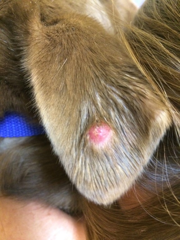
The small bump in the picture below is a small mast cell tumor. As you can see, it looks harmless and could easily be mistaken for a sebaceous cyst or lipoma. But don’t let its small size fool you: small tumors can be high-grade with the potential to spread quickly.
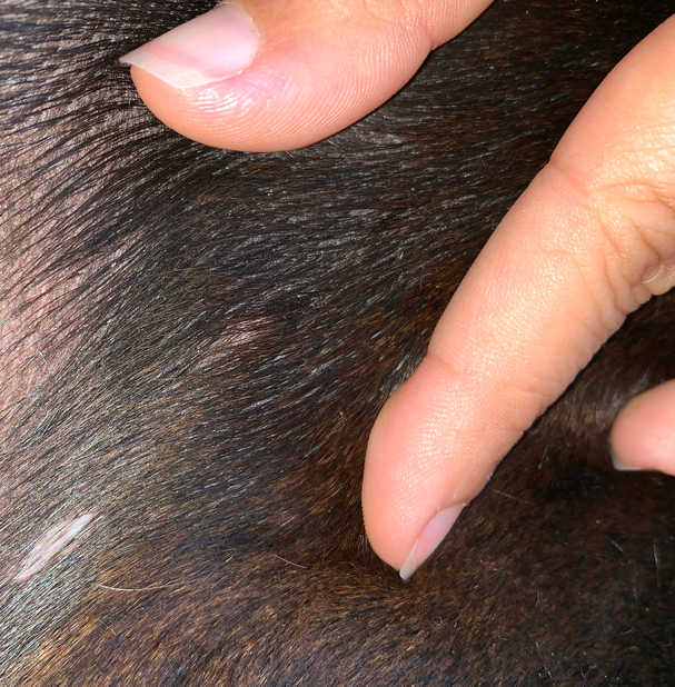
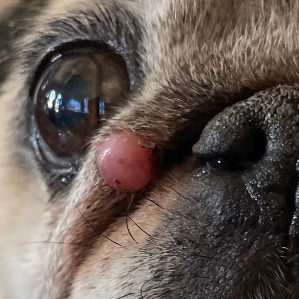
Small mast cell tumors can appear:
- Red or flesh colored
- Raised or flat
- Ulcerated
Small tumors can feel:
- Hard or soft
- Fixed or mobile
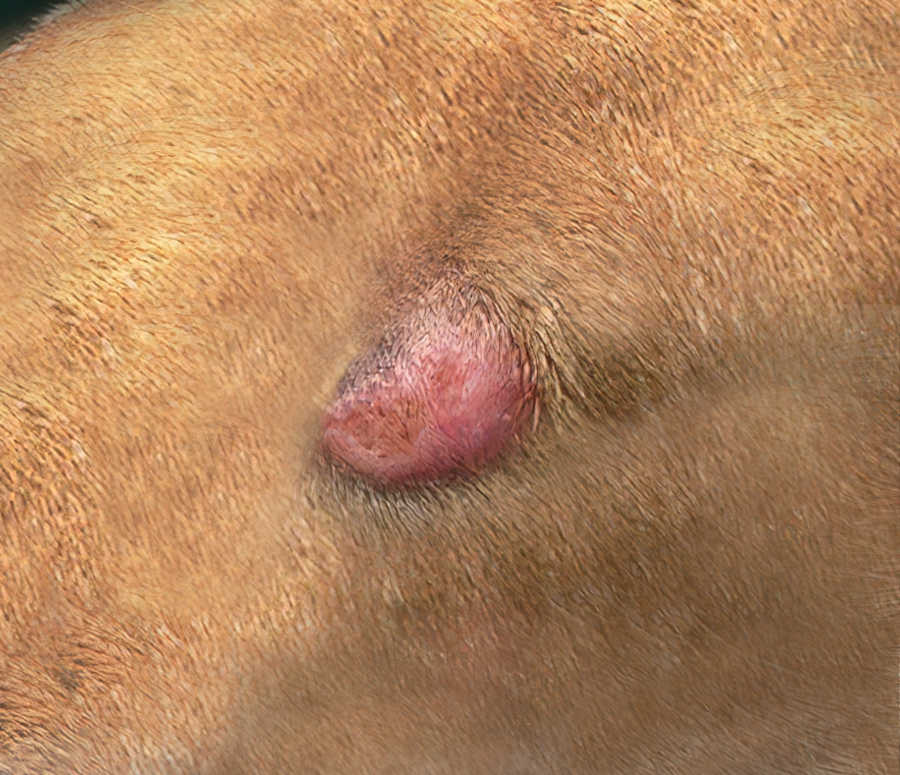
Large mast cell tumors
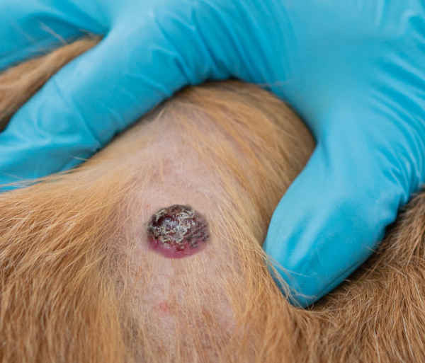
Below is a Pit Bull mix with a large mast cell tumor. The tumor appears damaged from the dog chewing and licking it. Tumors of this size are usually cancerous, high-grade, and at risk for metastasis (spreading). High-grade tumors need protection. A great way to prevent further damage is to put an e-collar on your dog. E-collars block the dog’s ability to reach the area and cause damage.
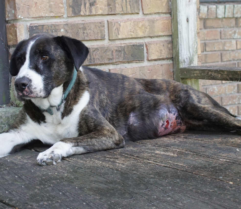
Large mast cell tumors look like:
- Large lumps
- Ulcerated skin on or around lumps
- Bleeding
- Itching
- Oozing skin
Large high-grade tumors can cause:
- Nausea
- Vomiting
- Weakness
- Diarrhea
2. Pictures by location
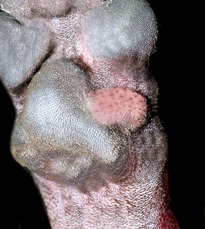
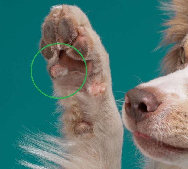
On a dog’s paw
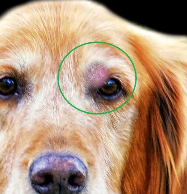
On a dog’s face or eyelid
Mast cell tumors near the eye are usually treated locally. However, they can spread to other parts of the body. Pictured below is a tumor on the eyelid of a senior Golden Retriever.
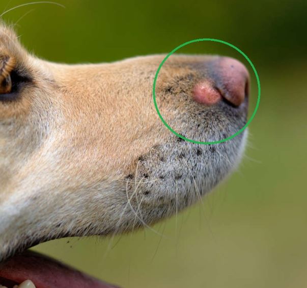
On a dog’s nose
Mast cell tumors on the muzzle (nose) are usually aggressive. These tumors have an increased risk of spreading.
Is it a mast cell tumor or something else? (2 examples)
Keep in mind that it is usually not possible to diagnose a tumor or growth just by looking at it: your veterinarian will likely want to do an FNA or biopsy to confirm diagnosis.
Example 1: is this a cancerous mast cell tumor?
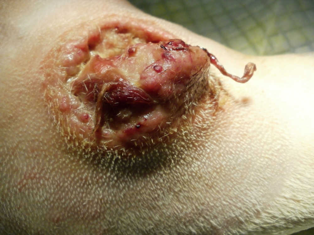
The picture below shows a possible cancerous growth that has ulcerated and is very inflamed:
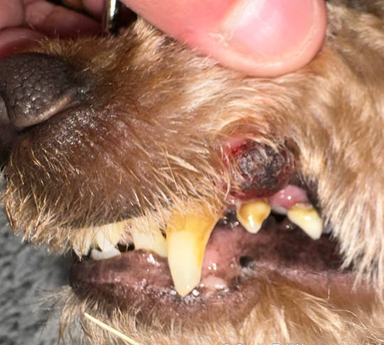
The skin appears to be necrotic (dead) and considerations for tumor type would include a melanoma, mast cell tumor or squamous cell carcinoma.
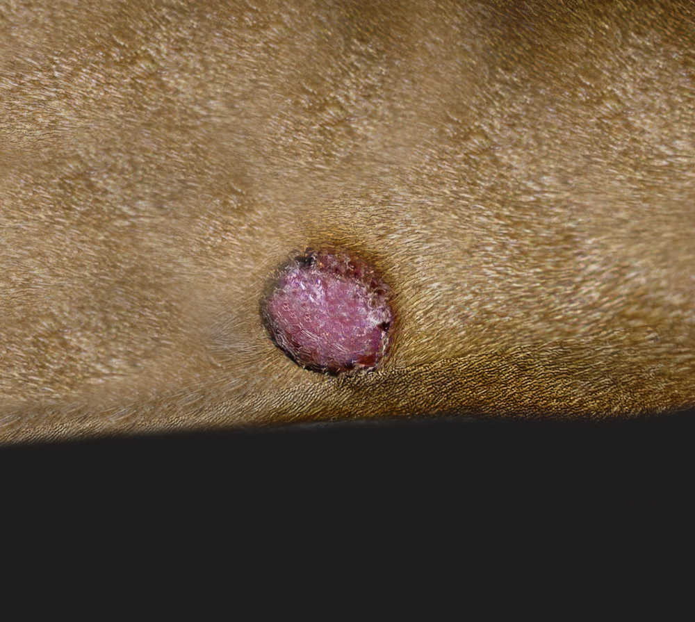
Example 2: Is this a mast cell tumor, or a benign wart or skin tag?
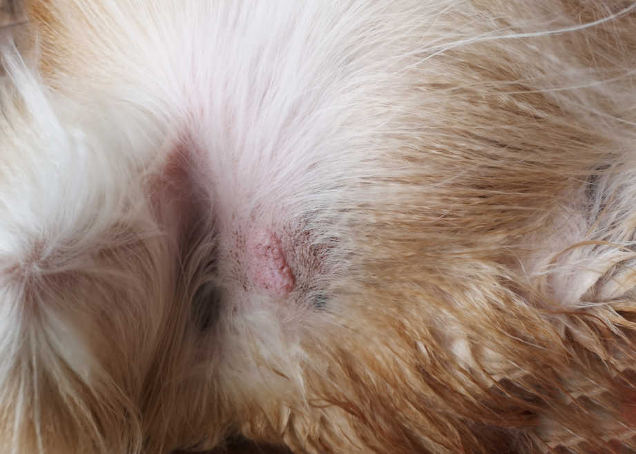
The picture below shows a pink and raised bump that is scabbed over and ulcerated in some areas. There also appears to be some flaked skin or keratin:
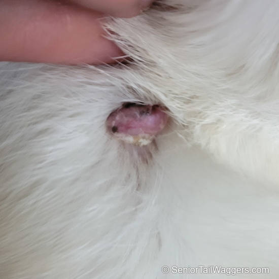
The growth on the picture above could be a benign wart or skin tag, but given how the surface is ulcerated, we should be concerned that there could be something more sinister going on here (such as a mast cell tumor or other kind of cancerous tumor). The only way to confirm diagnosis for sure is to sample it.
Example 3: is this a mast cell tumor or a histiocytoma?
A mast cell tumor is a cancerous tumor that forms when mast cells (immune cells) over-replicate. They come in all shapes and forms, but can sometimes look like histiocytomas (read our article: histiocytoma or mast cell tumor?). Veterinarians are usually not able to diagnose a lump or bump just by looking at it: diagnostic tests are often required to confirm diagnosis and determine the right treatment.
Related article: Pictures of the most common types of lumps and bumps in dogs.
Pictures of the surgery to remove mast cell tumors
While no two dogs will have the same surgery and recovery, knowing what to expect when your dog comes home from surgery is helpful. Here are some photos of what it may look like before, during, and after surgery.
Pre-surgical marking
In the image below, a veterinarian marked the skin around a mast cell tumor using a black surgical marker.
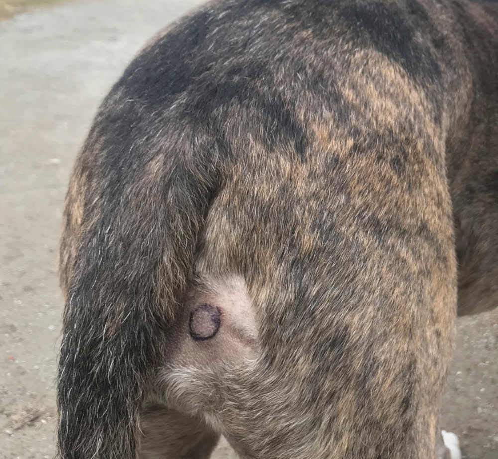
The mark is a guide to tell the surgeon where to cut during surgery. During surgery, the goal is to remove wide margins of skin around the tumor, decreasing the chance of tumor re-growth.
Your vet will also recommend a pre-surgical screening. These include a physical exam, blood work, and a surgical consultation. During the consultation, your veterinarian will explain the plan for surgery and answer any questions you have.
Recovering from surgery
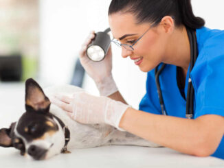
The dog in the photo below is getting his sutures checked. Sutures are typically removed 10-14 days after surgery, but large incision sites and ones in odd places may take longer to heal.
Sutures need to be clean and dry to prevent infection. Your dog may benefit from an e-collar or wrap to prevent them from licking the incision during the healing process.
Licking and chewing are the most common reason for suture failure, so watch your dog closely during this time. If you have an e-collar you can remove it when your dog eats and drinks, but make sure you watch them. All it takes is seconds for a dog to chew its incision open.
Picture of a dog with a wrap on his leg after mast cell tumor surgery
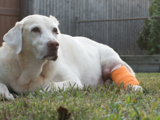
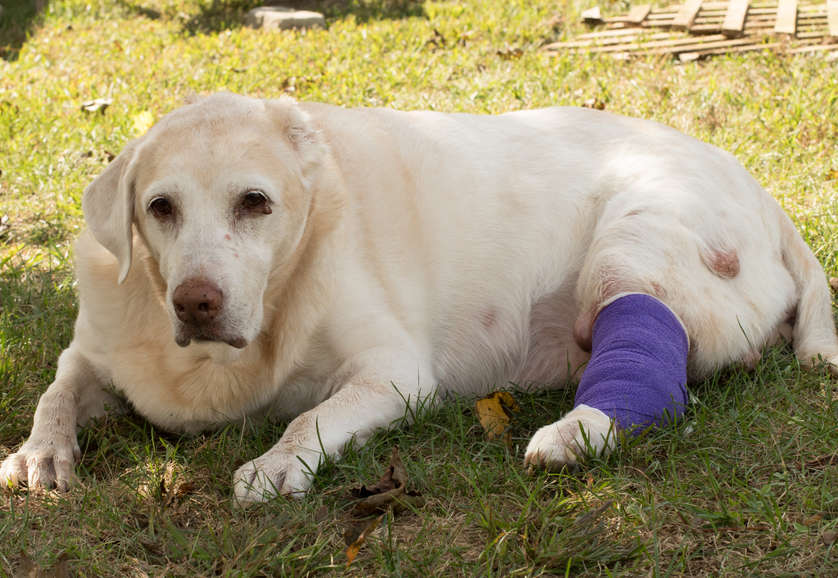
Each dog will have a different recovery. The length of recovery depends on the severity of the tumor. Dogs with higher-grade tumors may have a tough time recovering, while dogs with low-grade tumors may recover quickly. During recovery from surgery, wraps should stay clean, dry, and intact. Dirty, wet, or worn bandages can cause secondary infections. In the picture below, a senior Labrador Retriever is recovering from mast cell tumor surgery. His back leg has a wrap to prevent licking and protect it from damage and bacteria. Your dog may not have a wrap, depending on where the tumor is.
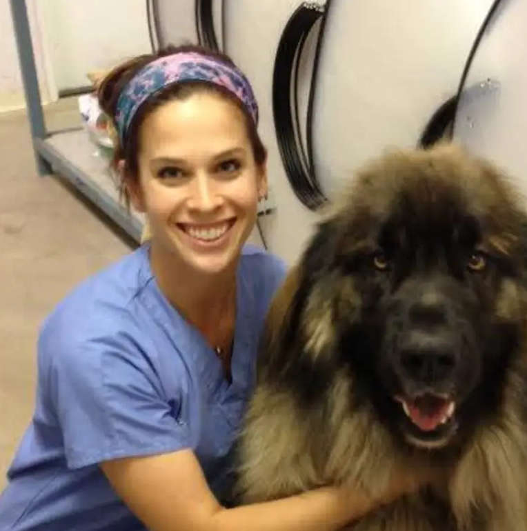
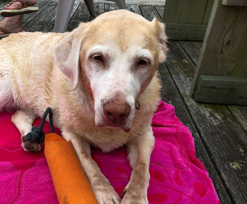
Below is a picture showing a senior Labrador drying off after a swim. The dog lost her ear to a mast cell tumor and is a courageous survivor!
More information about recovery after surgery
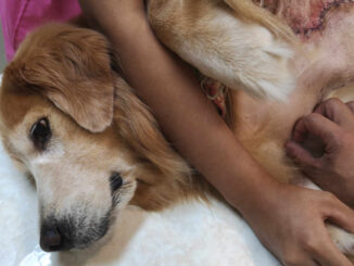
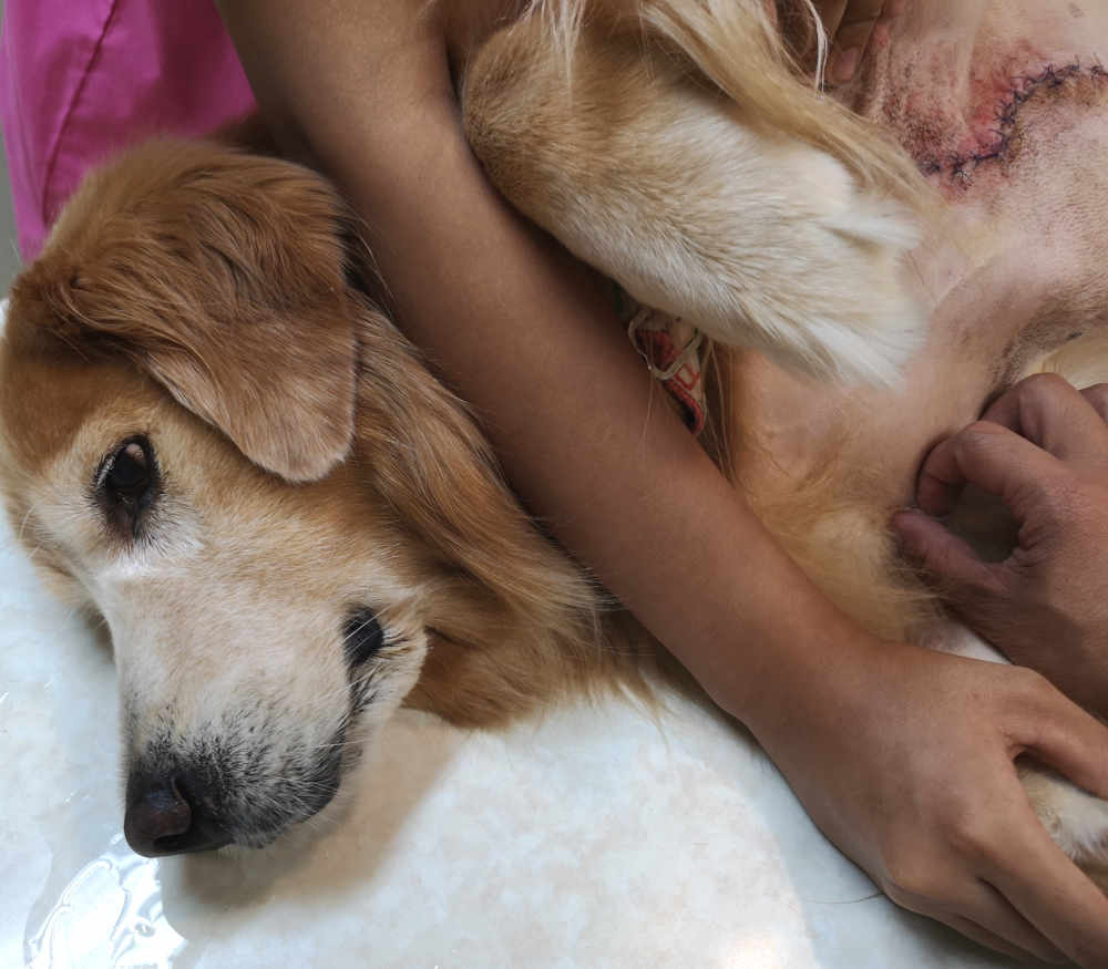
After surgery, your dog will have a recovery period before they wake up from anesthesia. The veterinarian will monitor their vital signs and ensure the incision is clean and protected. The dog in the photo below is in the process of waking up from a tumor removal surgery. After removal, the entire tumor, including the skin around it, goes out for biopsy.
At the lab, a pathologist will examine the edges of the sample for cancerous tissue. If the tissue sample shows cancer cells around the edges, your vet will recommend another surgery. Most tumors that return are high-grade and at risk of spreading. In these cases, veterinarians recommend a combination of medications, chemotherapy, and radiation. If your dog has symptoms related to the tumor, your vet may prescribe medications.
The most common medications used for dogs with mast cell tumors are:
- Steroids
- Anti-nausea medication
- Allergy medication
Your vet may also send your dog home with devices or aids to help during healing like:
- An E-collar (Elizabethan Collar)
- Wrap
- Bandage
Throughout the healing process, your dog will need periodic exams. During the exam, the veterinarian will check for the following:
- Swelling
- Infection
- Bleeding
- Failed or ripped sutures
- Any sign that the tumor may be coming back.
Life expectancy:
The life expectancy of a dog with a mast cell tumor varies and depends on the grade, stage, and location. Dogs with tumors in areas that are hard to treat (mouth, eyelid, and groin) will have the worst prognosis. For these dogs, surgery is not an option. They will benefit from chemotherapy, radiation, and medication. Out of the dogs who have low-grade tumors, 90-100% of them never come back. Dogs with high-grade tumors have a median survival rate of only six months after surgery. Learn more.
Picture of the rupture of a Mast Cell Tumor
Below is a photo of a mast cell tumor chewed open by a dog. Mast cell tumors release a substance called histamine that causes itchy skin. Some dogs itch so badly that they end up chewing their skin raw and creating a wound. The best way to prevent tumor rupture is by covering it with a clean wrap or putting an e-collar on your dog. To learn more about what to do when a mast cell tumor ruptures, read our article: Dog’s Mast Cell Tumor Itches, Bleeds or Bursts.
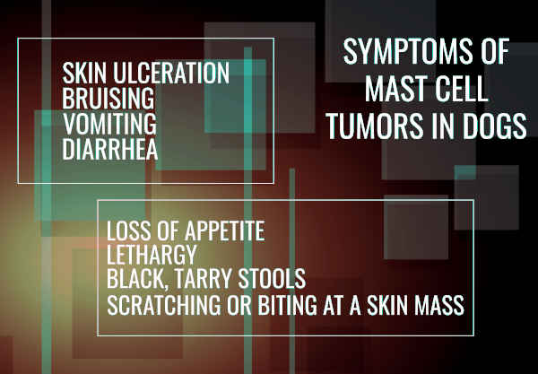
Early signs of Mast cell tumors in dogs
Because mast cell tumors look like many other skin conditions, it is hard to narrow the symptoms down. These tumors can look and act like ANY other skin condition! However, mast cells have one characteristic symptom that sets them apart from other skin conditions; they tend to change in size and appearance and grow larger or smaller from one day to another.
The earliest symptoms include:
- Lump or lumps that are itchy, or not
- Enlarged lymph nodes
- Irritated skin
Symptoms of more progressed mast cell tumors are:
- Welts
- Vomiting
- Diarrhea
- Weight loss
- Bloody or black, tar-like feces
- Abdominal pain
- Collapse
Learn more about mast cell tumors:
 Mast Cell Tumors vs Histiocytomas in Dogs [10 pictures] - According to research, histiocytomas make up 15.9% of tumors found in dogs, while mast cell… [...]
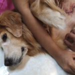 The Stages of Mast Cell Tumors and Life Expectancy [With Pictures] - When you find a new lump on your dog, it’s easy to dismiss it as… [...]
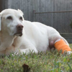 Mast Cell Tumors In Dogs: A Veterinarian’s Guide for Owners - Mast cell tumors are one of the most common forms of cancer in dogs. They… [...] What is My Dog’s Life Expectancy With Mast Cell Tumor? A Vet Weighs In - Just this past week, I cared for a dog who was recovering from a large… [...] What to Do When a Dog’s Mast Cell Tumor Itches, Bleeds, or Bursts - Mast cell tumors are a common skin cancer in dogs, accounting for 20% of all… [...]
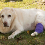 When to Euthanize Your Dog with Mast Cell Tumors [Veterinarian Advice] - The worst part about owning a dog is that their lives are far too short.… [...]
Author
- Bostock, D.E. (1986). The prognosis following surgical removal of mastocytomas in dogs. Journal of Small Animal Practice, 27(10), 723-737. [↩]
Disclaimer: This website's content is not a substitute for veterinary care. Always consult with your veterinarian for healthcare decisions. Read More.