

This article was updated on October 9th, 2023
As a veterinarian, I often come across dog parents who are concerned about their pet’s skin lesions or lumps and immediately assume that it could be cancer. However, not all skin lesions or lumps are malignant and many of them are usually harmless. Nonetheless, it is crucial to differentiate between benign and cancerous lumps.
In this article, our veterinarians explain the characteristics of skin cancer and cancerous lumps, along with example pictures.
5 Characteristics of Cancerous Lumps or Lesions
Cancerous, or malignant, skin lesions and tumors can be small or large. They may itch or cause the senior dog some discomfort. They may do neither. You can never say whether they are cancerous just by looking at them, but there are some clues that can help raise suspicions:
1. Cancerous lumps or lesions are often growing rapidly
Cancer is the abnormal and rapid growth of previously healthy cells. Therefore, lesions or lumps that are growing rapidly or changing in appearance quickly may indicate a more sinister underlying cause. If they double in size over the course of a few weeks, it is best to get them checked as soon as possible.
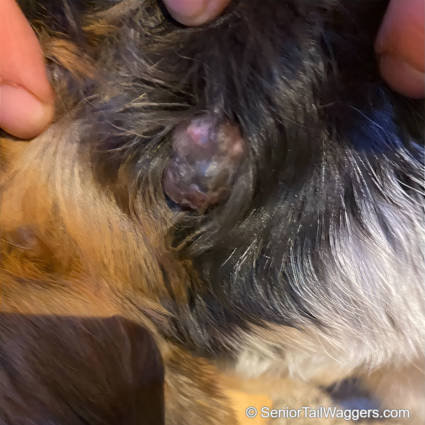
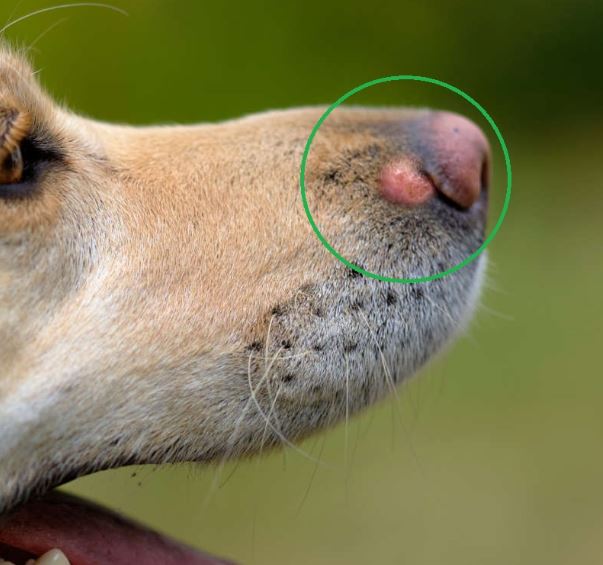
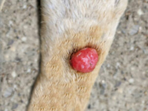
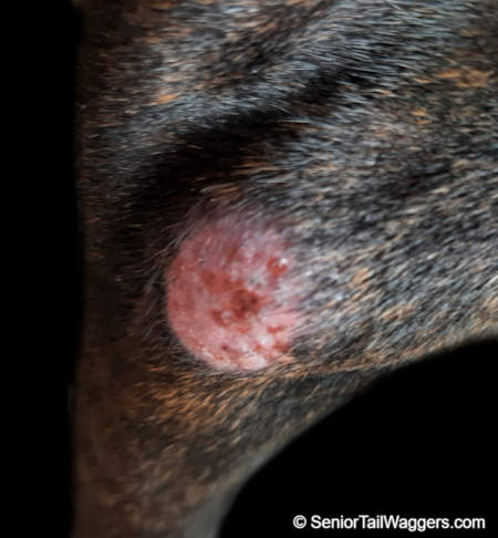
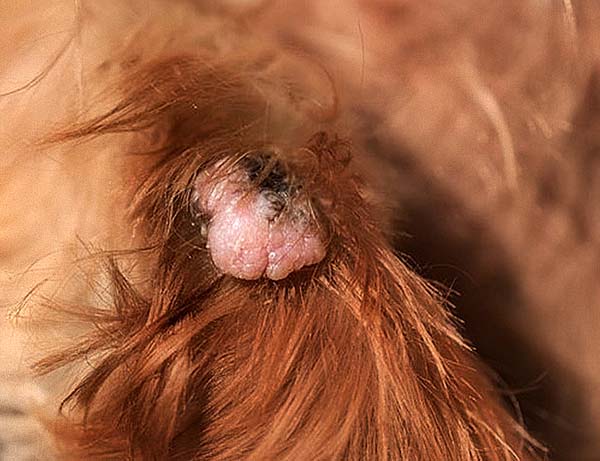
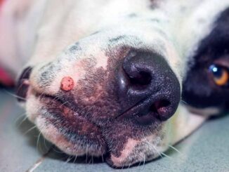
The picture below shows a senior dog which has developed a raised, irregular and fleshy growth. The growth contains a mixture of pigments (black/pink). Because this mass has grown rapidly, there is an increased risk that the growth could be cancerous:
2. Cancerous lumps are often harder and firmer
Cancerous lesions and lumps are often harder and firmer to the touch. Ulceration, redness, and a firm texture are all common properties, but that doesn’t mean that every lump with these features will be malignant.
3. Cancerous lumps tend to have more irregular shapes
Due to their rapid and erratic growth, cancerous lumps tend to appear more irregular in shape. Red, black, or just generally ‘unhealthy’ looking lumps or lesions may be more malignant in origin.
4. Discharge may occur
Oozing or discharge from the lesion may occur due to damage and death of the tissue in surrounding areas. While any lump can develop a secondary infection, sinister lumps are more prone to producing pus and bleeding.
5. Cancerous lumps or lesions tend to cause more discomfort
A lump that is itchy or causing discomfort to your dog is more likely to be malignant.
“It’s often not possible to determine the true nature of a lump simply by looking at it. A lump that appears harmless could potentially be cancerous, and vice versa. To obtain an accurate diagnosis and determine the appropriate treatment, a Fine Needle Aspirate or biopsy is necessary to confirm the diagnosis.”

Learn more about the vet process and costs to diagnose lumps and bumps, including Fine Needle Aspirates and Biopsies.
Pictures of Skin Lesions or Lumps that Are Cancerous (or Often Cancerous)
1. Mast Cell Tumors
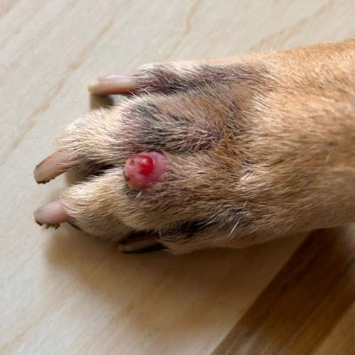
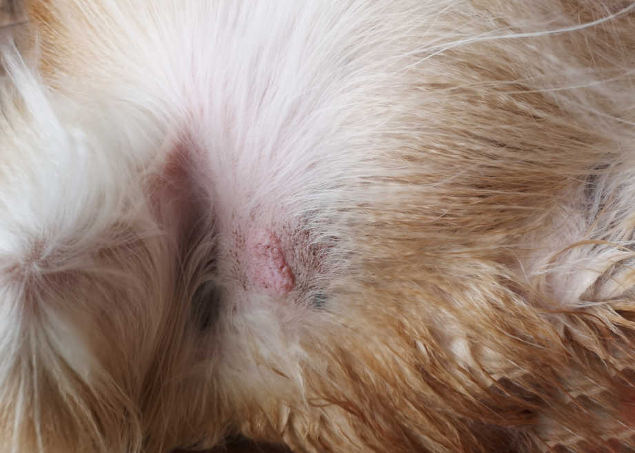
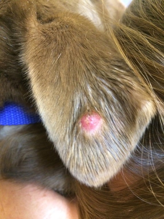
Mast cell tumors are the most common form of skin cancer in dogs. Mast cell tumors come in many shapes and sizes. They are more often seen in middle-aged and older dogs but can affect younger dogs too. Below are several pictures showing mast cell tumors:
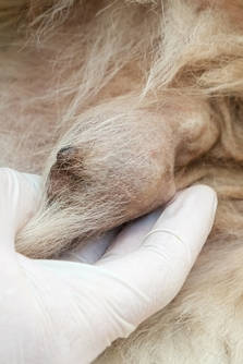
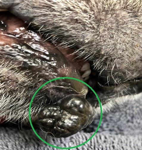
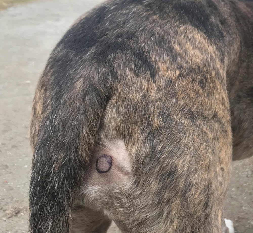
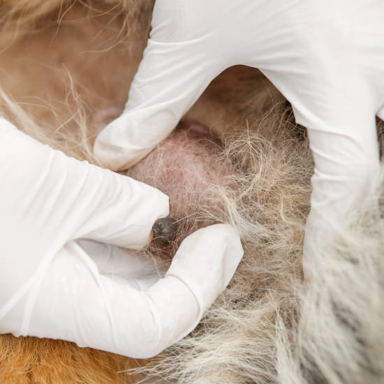
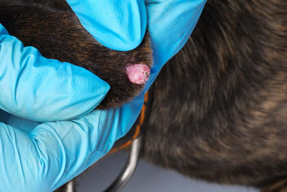
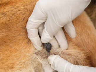
These old dog lumps are not always malignant, but it’s impossible to know which ones are and which aren’t without a fine needle aspirate or biopsy. Pictured below is another cancerous mast cell tumor on a dog (circled with a pen before surgery to remove the tumor):
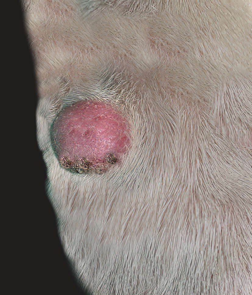
Mast cell tumors can vary a lot in appearance; usually, they’re a smooth, round growth visible on the skin. Other times they can look like a wart, or resemble a lipoma, and sometimes they are red in color.
Learn more about Mast Cell Tumors (with pictures & advice from our veterinarian team).
2. Melanomas
Melanoma tumors are dark and can be small, large, flat, or raised. They can be either benign or malignant, so they shouldn’t be ignored. Most malignant melanomas in dogs grow in/around the mouth or in other mucus membranes, but they can also be found in other areas.
The pictures below also show melanomas (black growths) inside a dog’s mouth, on the lips and eyelids:

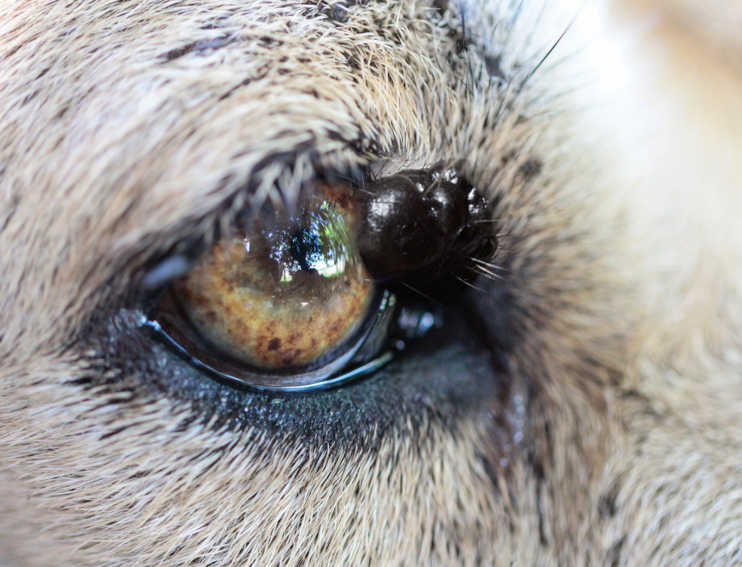
If a dog has malignant melanoma, it’s usually an aggressive cancer that spreads throughout the body quickly, so the tumor needs to be surgically removed as quickly as possible. Only a veterinarian can tell the difference between a benign and a malignant melanoma by doing a biopsy.
Learn more about Melanomas.
3. Squamous Cell Carcinomas
Squamous cell carcinomas in dogs are fairly rare, and they are not as aggressive in terms of spreading as melanoma or mast cell tumors are.
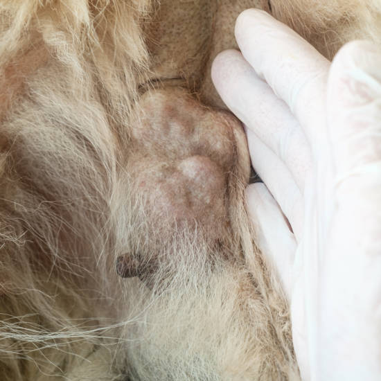
These types of malignant tumors are usually found on areas of skin that are bare, or have little hair, and are more common in dogs with light-colored skin. They are almost always on the belly and inner thighs (areas of thin or no hair), but can appear anywhere.
UV radiation from the sun can contribute to the development of these tumors. Squamous cell tumors in dogs can be raised lumps or nodules, or flatter areas of ulcerated skin. They can sometimes resemble warts.
4. Mammary Gland Tumors
Mammary gland tumors can be cancerous or benign. They are often located next to or beneath the nipple and may extend between multiple mammary glands. They are firm and may have ulcerated skin overlaying or be abscessed and bleeding.
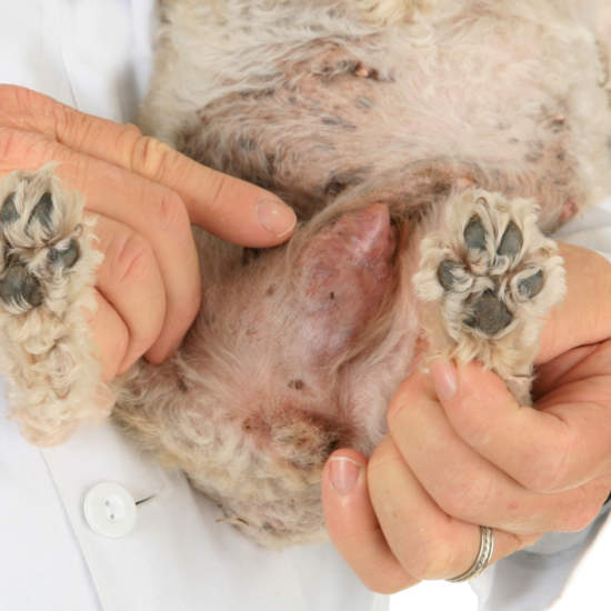
Due to the lymphatic drainage that links mammary glands together, these tumors can spread quickly to the other mammary glands and even to the rest of the body. Therefore, quick surgical removal is recommended.
Learn more about Mammary Gland Tumors.
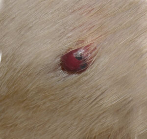
5. Sarcoma Tumors
Hemangiosarcoma is a malignant cancer of the cells that line the blood vessels of the body. Therefore, hemangiosarcoma can occur anywhere in the body where blood vessels are found. In the case of hemangiosarcoma, the cells that line the blood vessels start dividing uncontrollably and in an unhealthy manner. This leads to the development of a mass that is very prone to rupture and can even cause a dog to bleed out internally.
Read more about Hemangiosarcoma.
Skin Issues or Lumps That Are Mostly Benign (Non-Cancerous)
Not all skin growths are cancerous – in fact many are nothing to worry about, but should still be checked out by your veterinarian. Below are several examples of bumps often seen on dogs that are mostly benign:
1. Warts. Warts are mostly benign and can be left alone in most cases. Occasionally, warts can become cancerous so any wart that is a long-term issue, or that changes in color/size/look needs to be investigated by a vet. Learn more or see images below for example warts on dogs:
2. Histiocytomas: They can look pretty scary to the untrained eye, and it can be hard to tell them apart from dangerous growths in dogs. However, a histiocytoma is a benign (non-cancerous) growth, often found on the skin of usually young dogs. These growths form when a type of immune cell (histiocyte) in your dog’s skin over-replicates. Most histiocytomas will heal without treatment in 2-3 months. Learn more.
Other mostly benign skin issues include skin tags or sebaceous adenomas. For more information, check out our article featuring Common Skin Lesions in Dogs.
What to Do if You Find a Suspicious Skin Lesion on Your Dog
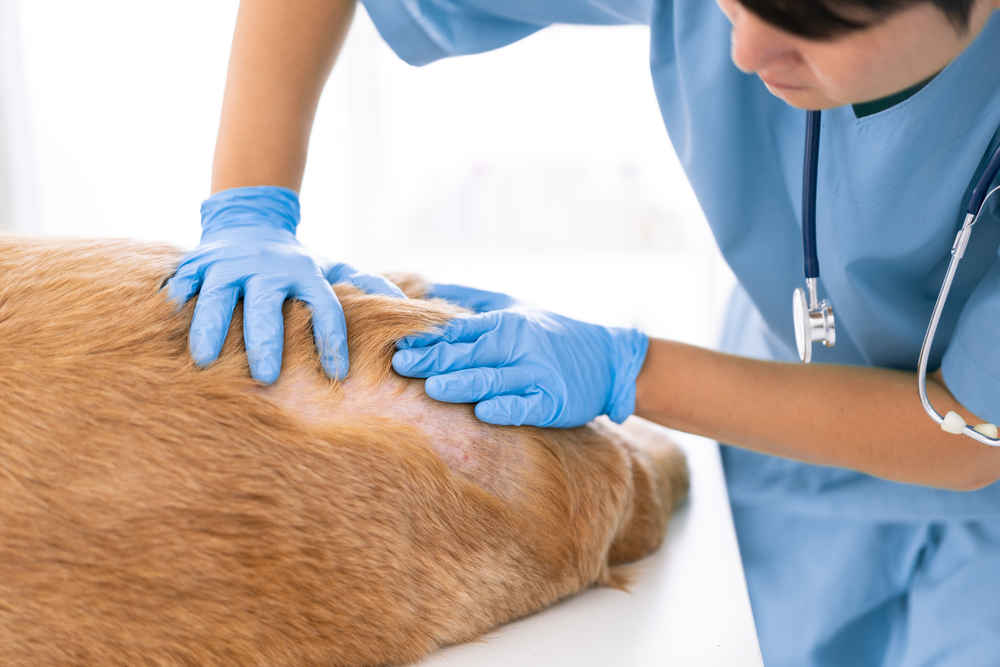
Regularly check your dog for changes in their skin. Run your hands through their coat once a week, and be sure to feel in all the creases and folds for any abnormal lumps or lesions. Make a note of the size and the general appearance – a photograph can help you keep a diary in this way.
So, what happens if you find a skin lesion? There are two options: monitor it for a while and see if it changes or take your dog to the vet for a checkup. We would always recommend the latter if you’re not sure, particularly if it looks sinister. We will discuss more on how to tell a cancerous lump from a benign one next.
Diagnosis and treatment
As mentioned earlier, it is usually not possible to confirm diagnosis just by looking at a lump. The diagnosis will involve your vet taking samples and analyzing them under a microscope in a process known as histopathology. Histopathology is performed by a board-certified veterinary pathologist in a laboratory so the sample will need to be sent in for analysis.
Different sampling techniques can be performed including:
- Fine needle aspirate – a needle with a syringe attached is inserted into the lesion, and a sample is taken by pulling back on the plunger of the syringe. This aspirates some cells that can be placed on a microscope slide for analysis. The advantage of this technique is that many dogs will tolerate having it done consciously. However, it won’t always achieve a diagnostic sample as only a pinprick sample is taken. Furthermore, not all lumps will readily release cells for aspiration, so this technique doesn’t always work.
- Biopsy – a biopsy is a wedge of tissue that is taken from the lesion and then sent for analysis. This is performed under a general anesthetic and is the gold standard for getting a diagnosis of a skin lesion. Often the edge of the lesion will be taken to allow the histopathologist to compare normal to abnormal tissue.
Learn more about fine needle aspirates and biopsies.
Treating Malignant Skin Tumors in Dogs
The best way to treat most malignant or cancerous lumps in dogs is to remove them surgically, and as quickly as possible. The smaller the lump the easier the surgery and the less tissue which must be removed. A malignant lump will often be removed with a margin of normal-looking tissue around it – this is to ensure that any microscopic spread of the cancer is also removed.
Acting fast also helps to reduce the chances of the cancerous cells metastasizing (traveling) into other tissues, organs, and lymph nodes. Sometimes radiation therapy, chemotherapy, or other treatments are recommended in addition to removing the tumor. Your vet may wish to refer your dog to an oncologist to aid in this treatment. There are other treatments, such as injectable medications for tumor removal as well as cancer vaccines.
The exact treatment options will be decided by your vet and consider the size and location of the tumor and whether it has spread to other areas of the body.
Related Posts:
- Common Skin Lesions in Dogs
- Skin Cancer in Dogs
- What Steps can be Taken to Prevent Canine Skin Cancer?
- How Long Can dogs live with Skin Cancer?
- What is the Best Way to Care for my Dog who has Skin Cancer?
Author
Disclaimer: This website's content is not a substitute for veterinary care. Always consult with your veterinarian for healthcare decisions. Read More.
We have a 12-year-old mastiff/ lab mix and she has a huge red growth on the outside of her upper lip. Just like the first photo but on the lip. It is quite large. Is that a cancerous tumor?
These lumps or growths are not necessarily malignant. However, you should get your dog checked out by a vet as they will be able to do further testing to determine the true nature of the growth. It’s really challenging to make an accurate diagnosis just by looking at a lump. If your dog needs treatment, the sooner you act, the better.
A dog was basically dropped off at my driveway, seemed in good health, not underweight, no fleas and a healthy appetite. After further inspection I saw a soft/firm or slightly firm growth on on leg above paw. Hairless,painless and about the size of a charry tomato, a large cherry tomatoe but still about that size. Doesn’t seem to cause pain. We have an appointment On Monday after the weekend. Hoping it isn’t cancer. Can’t find anything online that looks like what he has. I have been looking for a dog for soo long. And was so excited to see one just walk right into my life. Fingers crossed it will be something treatable.