
This article was updated on August 3rd, 2023
Dogs are predisposed to a variety of skin conditions that can cause cysts, lumps and bumps to form. In this article, we will review 4 common cysts often found on dogs’ tails, and what to do about them. At the end, we will also review common tail lumps that can be mistaken for cysts.
My dog has a cyst on its tail – what could it be?
Different varieties of cysts can develop on a dog’s tail. These are some examples:
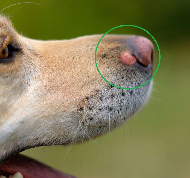
- Sebaceous Cysts: These are the most common types of cysts in dogs. They occur when the sebaceous glands, which produce oil to lubricate the skin and hair, become blocked or damaged. Sebaceous cysts are usually benign and may appear as small, firm bumps under the skin.
- Follicular Cysts: These cysts can form when a hair follicle becomes blocked or damaged. They often appear as fluid-filled sacs and can range in size.
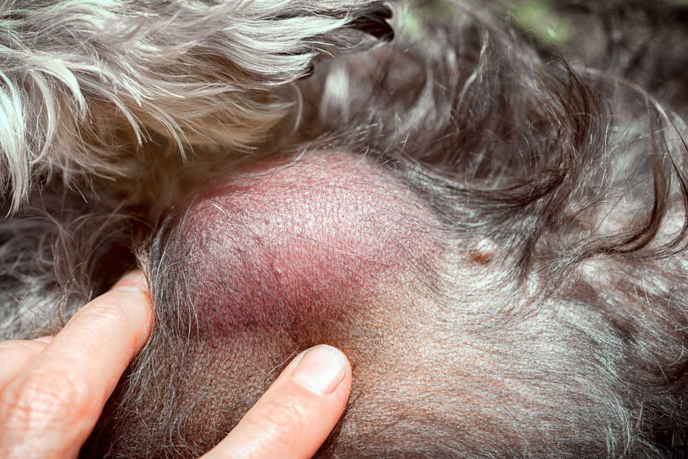
- Pilonidal Cysts: These cysts are typically found at the base of the tail and are more common in certain breeds. Pilonidal cysts can result from hair follicle infections or ingrown hairs and may become painful or infected.
- Abscesses: An abscess is a localized infection that can develop anywhere on the body, including the tail. Abscesses typically appear as a swollen, painful lump filled with pus.
Related post: Lumps Often Found on a Dog’s Tail
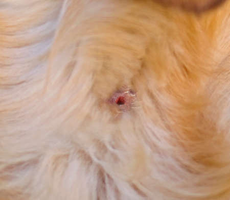
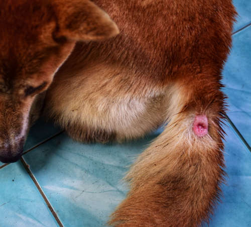
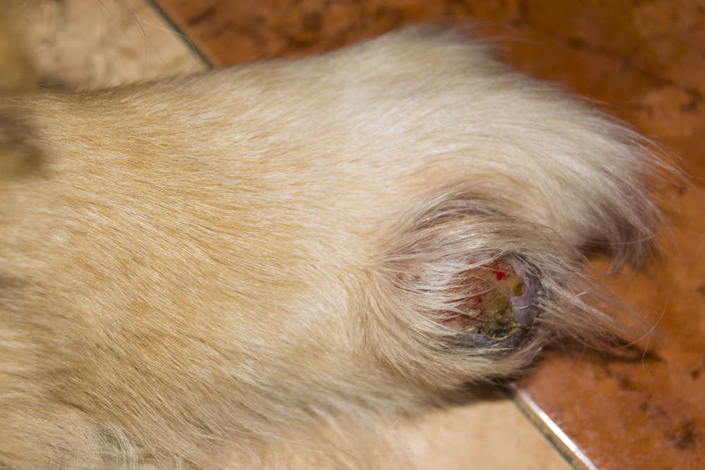
4 types of cysts often found on a dog’s tail [with pictures]
Now that we discovered the various types of cysts, let’s get into more detail about each of these cysts.
1. Sebaceous Cysts:
A sebaceous cyst on a dog is a common skin condition characterized by the formation of a benign, fluid-filled sac beneath the skin. These cysts form when sebaceous gland ducts become blocked. The sebum produced by these glands starts to accumulate and eventually forms a cyst.
Key characteristics of sebaceous cysts:
- small, round, or oval-shaped bumps under the skin
- size varies from a few millimeters to several centimeters in diameter
- typically smooth, firm to the touch, and may be movable under the skin
While sebaceous cysts are usually harmless, they can occasionally become inflamed, infected, or rupture. Signs of an infected sebaceous cyst may include redness, swelling, pain, discharge, or a foul odor.
In most cases, sebaceous cysts on dogs do not require treatment unless they become bothersome or infected. If the cyst causes discomfort, interferes with movement, or poses a risk of infection, your veterinarian may recommend surgical removal.
Learn more about sebaceous cysts in dogs.
2. Follicular Cysts:
A follicular cyst forms when a hair follicle becomes blocked or damaged. Hair follicles are the structures from which hair grows. When the normal process of hair growth is disrupted, a follicular cyst can develop.
Follicular cysts in dogs often appear as fluid-filled sacs under the skin. They can vary in size and may feel soft or firm to the touch. These cysts are typically round or oval-shaped and may be located anywhere on the dog’s body where there are hair follicles.
Follicular cysts can be caused by various factors, including hormonal imbalances, genetic predisposition, or skin inflammation. Some dog breeds may be more prone to developing follicular cysts than others.
While follicular cysts are generally benign and not typically painful, they may cause cosmetic concerns or discomfort if they become large or inflamed. Treatment may involve draining the cyst, administering medications to address any underlying causes, or, in some cases, surgically removing the cyst.
Learn more about follicular cysts in dogs.
Related post: Lumps Often Found on a Dog’s Tail
3. Pilonidal Cysts:
Pilonidal cysts are a specific type of cyst that typically occur in the sacrococcygeal area, which is the region near the base of the spine above the tailbone. Pilonidal cysts are relatively rare in canine patients.
A pilonidal cyst in dogs is characterized by the formation of a cyst or abscess in the skin and subcutaneous tissue of the sacrococcygeal area. The exact cause of pilonidal cysts is not fully understood, but it is believed to involve hair follicle infections or ingrown hairs in the area. When hair penetrates the skin and triggers an inflammatory response, it can lead to the development of a cyst or abscess.
Symptoms of a pilonidal cyst in dogs may include:
- swelling,
- pain,
- redness,
- discharge, and
- the presence of a visible lump or abscess in the sacrococcygeal region.
The cyst or abscess can become infected, resulting in additional symptoms such as fever, increased pain, and systemic signs of illness.
Treatment for pilonidal cysts in dogs typically involves a combination of medical management and surgical intervention. Your veterinarian may prescribe antibiotics to address any infection present and provide pain relief. In some cases, the cyst or abscess may need to be drained or surgically excised under general anesthesia. Surgery aims to remove the cyst and any associated infected tissue, allowing for proper healing.
4. Abscesses:
An abscess in dogs is a localized infection characterized by the collection of pus within a pocket or cavity. It typically occurs as a result of bacterial contamination or the body’s response to foreign material, such as a splinter, thorn, or bite wound.
Common signs and symptoms of an abscess in dogs include:
- Swelling: The affected area may appear swollen or enlarged.
- Pain: Dogs may exhibit signs of discomfort, such as limping or reluctance to move.
- Heat: The area around the abscess may feel warm to the touch.
- Redness: The skin surrounding the abscess can become reddened.
- Drainage: In some cases, the abscess may rupture, resulting in the release of pus or a bloody discharge.
Treatment for abscesses typically involves a combination of medical management and, if necessary, surgical intervention. The veterinarian may need to drain the abscess, clean the area, and prescribe antibiotics to combat the infection. In certain situations, the veterinarian may also need to surgically remove the infected tissue or address any underlying causes that contributed to the abscess formation.
Related post: Lumps Often Found on a Dog’s Tail
Is a cyst on a tail painful to the dog?
Whether a cyst on a dog’s tail causes pain can vary depending on several factors, including the size, location, and underlying cause of the cyst. In general, small and uncomplicated cysts may not cause any pain or discomfort to the dog. They can be relatively harmless and go unnoticed by the dog unless they grow larger, become inflamed, or become infected.
It’s important to monitor your dog’s behavior and look for signs of discomfort or pain, such as:
- licking or chewing at the cyst,
- reluctance to sit or lie down, or
- changes in activity levels.
If you notice any concerning symptoms or your dog appears to be in pain, it’s best to consult with a veterinarian for a proper evaluation and appropriate treatment options.
Veterinary treatments are recommended in case of infection, discomfort, or growth
In general, small and uncomplicated cysts that are not causing any discomfort or functional problems may not require immediate treatment. These cysts can be closely monitored to ensure they do not grow or become infected.
However, there are situations where treatment may be recommended:
- Infection: If the cyst becomes infected, it may require treatment to address the infection and prevent further complications. This may involve draining the cyst, cleaning the area, and potentially prescribing antibiotics.
- Pain or discomfort: If the cyst is causing pain, irritation, or interfering with your dog’s movement or quality of life, treatment may be necessary. This can involve surgical removal of the cyst to alleviate the symptoms.
- Rapid growth or size: If the cyst grows rapidly or becomes excessively large, it may be recommended to remove the cyst for diagnostic purposes or to relieve any potential issues it may cause.
- Behavioral changes: If you observe changes in your dog’s behavior, such as increased licking, chewing, or persistent scratching at the cyst, it may indicate that the cyst is bothering your dog. In such cases, it’s best to consult with your veterinarian to determine the cause and appropriate treatment options.
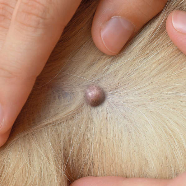
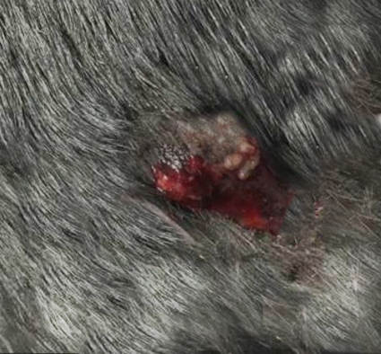
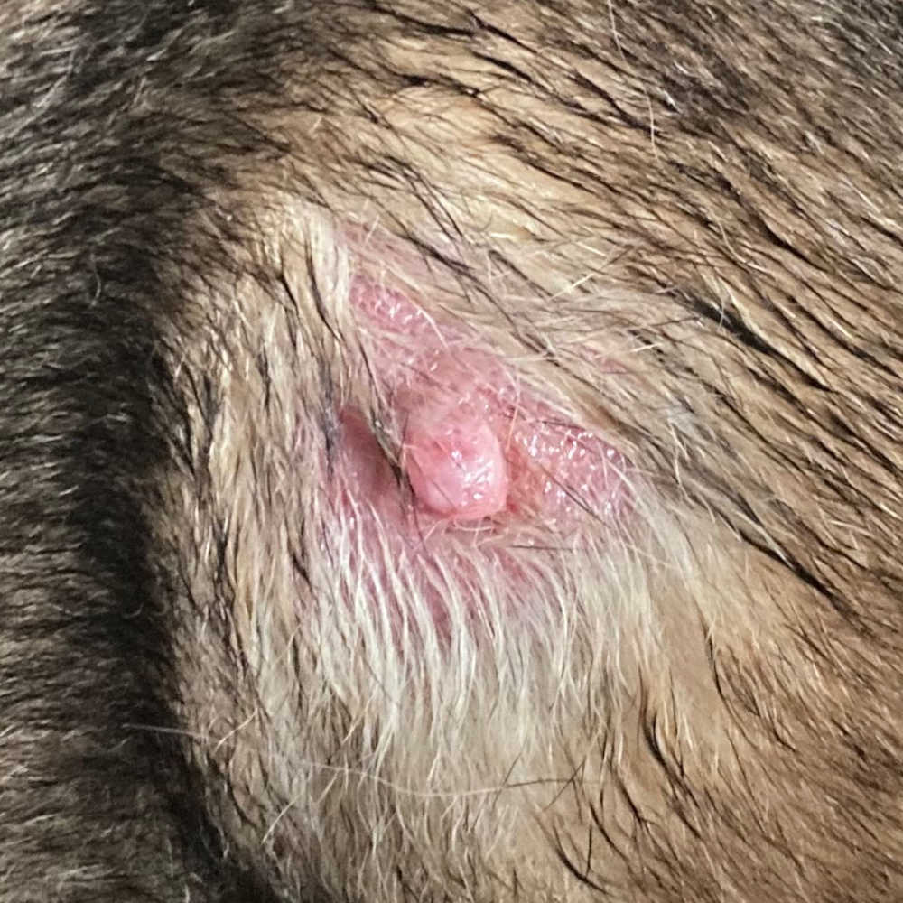
Below is a photo of a cyst that is ulcerated and infected by a dog chewing it. This cyst requires treatment to prevent further damage and worsening of infection:
Is there anything that I can do at home?
Here are a few general measures you can take at home to help manage a cyst on your dog’s tail:
- Keep the area clean: Gently clean the area around the cyst with a mild, pet-safe antiseptic solution or warm water and gentle soap. This can help prevent infection and maintain hygiene.
- Monitor the cyst: Keep an eye on the cyst for any changes in size, color, or discharge. Note any signs of discomfort or worsening symptoms.
- Prevent licking or chewing: If the dog is bothering the cyst by licking or chewing at it, you can use an Elizabethan collar (cone) to prevent access to the tail. Excessive licking or chewing can lead to irritation or introduce further infection.
- Do not attempt to drain or pop the cyst: It’s generally not recommended to attempt draining or popping the cyst at home. This can introduce further infection, cause trauma, and potentially worsen the condition.
Diagnosing your dog’s tail issues at the vet
When you take your dog to the veterinarian for a cyst or bump on their tail, the veterinarian will perform a thorough examination and may follow these steps to diagnose the condition:
- Physical examination: The veterinarian will physically examine the cyst and the surrounding area. They will note its size, shape, color, texture, and any signs of inflammation or infection. The veterinarian may also palpate the area to assess its consistency and mobility.
- Fine-needle aspiration (FNA): In many cases, the veterinarian may perform an FNA, which involves using a small needle and syringe to collect a sample of cells and fluid from the cyst. The sample is then examined under a microscope to determine the nature of the cyst and whether any abnormal cells or infections are present.
- Biopsy: In certain situations, the veterinarian may recommend a biopsy to obtain a larger tissue sample for analysis. This may involve surgically removing a portion of the cyst or performing an excisional biopsy to remove the entire cyst. The tissue sample is then sent to a laboratory for further examination.
Other types of bumps often found on a dog’s tail
It is usually hard to diagnose a lump, bump or cyst just by looking at it. To learn more about the types of bumps often found on a dog’s tail, read our veterinarian article: “New lump or bump on a dog’s tail: what is it?“. For example:
Mast cell tumors: Mast cell tumors can develop on any part of the body, including the tail. These tumors arise from mast cells, which are part of the immune system. Find out what mast cell tumors look like.
Warts: Warts, also known as papillomas, are benign growths caused by viral infections. They often appear as small, raised bumps on the skin, including the tail.
Hematomas: Hematomas are collections of blood that can occur due to trauma or injury. They can cause a localized swelling or lump on the tail and may be accompanied by pain or discomfort.
Tumors: Various types of tumors, both benign and malignant, can develop on a dog’s tail. These can include fibromas, fibrosarcomas, adenomas, or adenocarcinomas. The appearance, growth rate, and behavior of these tumors can vary. View more pictures of lumps and bumps.
Lipomas: While less common on the tail, lipomas are benign fatty tumors that can occur in dogs. They usually feel soft and moveable under the skin.
Related posts:
Author
Disclaimer: This website's content is not a substitute for veterinary care. Always consult with your veterinarian for healthcare decisions. Read More.