


This article was updated on August 24th, 2024

Seeing skin issues is a nearly everyday thing at our veterinary clinic. This article will help you identify lumps, bumps, or sores that are often found on dog paws, with the aid of pictures provided by our veterinarians. We will cover the most frequent causes, but it’s important to keep in mind that you should always consult your vet for proper diagnosis and treatment.
8 lumps, bumps or sores often found on dog paws
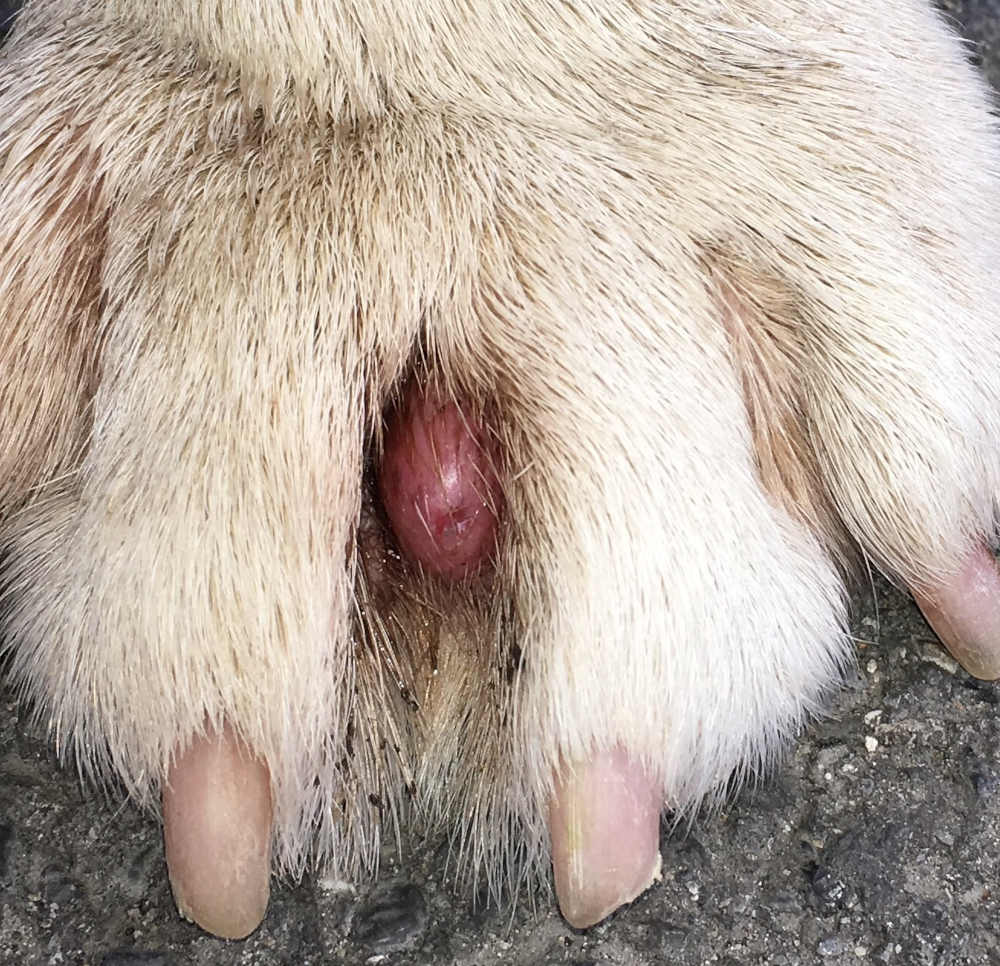
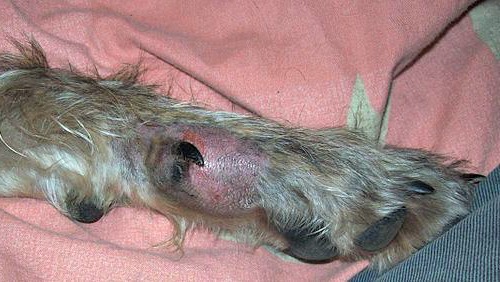
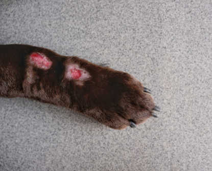
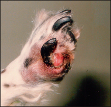
1. Interdigital cysts
These red bumps pop up between your dog’s toes and are often filled with blood or pus. They can become quite large, as shown on the picture below. If painful enough, your dog will either limp or lick them excessively. Treatment often involves an antibiotic and anti-inflammatory if they are causing a lot of swelling. Learn more about Interdigital Cysts.
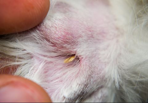
2. Foreign body
Your dog’s feet go almost everywhere! This gives grass seeds, thorns, and thistle stickers an easy route to get into them and cause problems. Once foreign bodies make their way into the skin between a dog’s toes or into the foot pad, they’re free to set up a nasty inflammatory reaction complete with redness, swelling, and heat.
These lumps or skin lesions will cause a dog to lick and chew their feet and may start to ooze thick, colored discharge. Without treatment, foreign bodies can cause abscesses or, in some cases, infections. To resolve these lumps or lesions, veterinary treatment is usually necessary. Neglecting an infection or foreign object can make the condition worse and increase the risk of complications.
Soaking your dog’s foot and getting antibiotics from your veterinarian will get rid of most foreign body issues. If foreign material is present, your vet can sometimes remove it with local anesthetic, but more thorough surgical exploration under sedation or general anesthetic may be necessary (estimated cost: from $50-$100 for a vet visit and antibiotics to $500 or more for surgical removal).
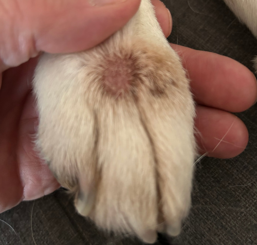
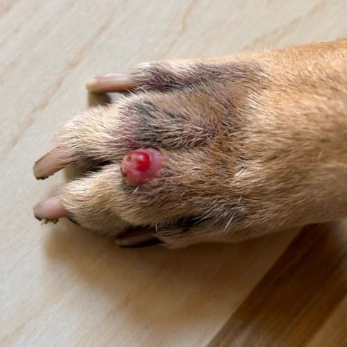
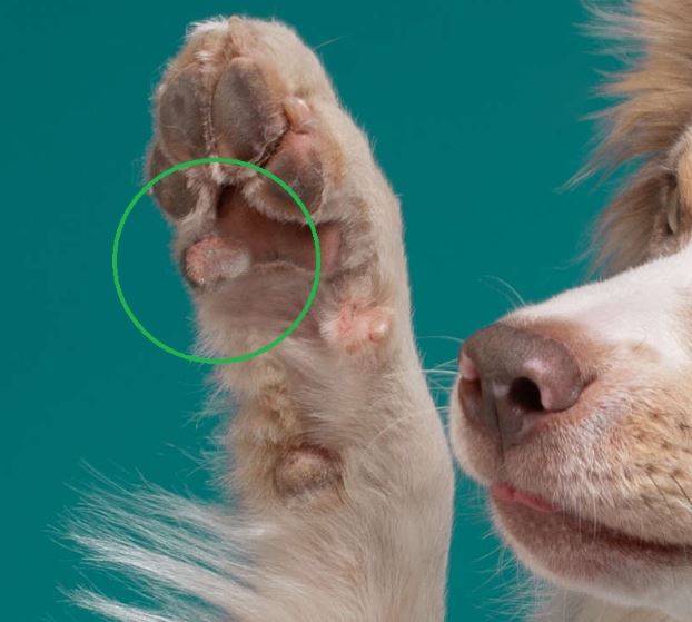
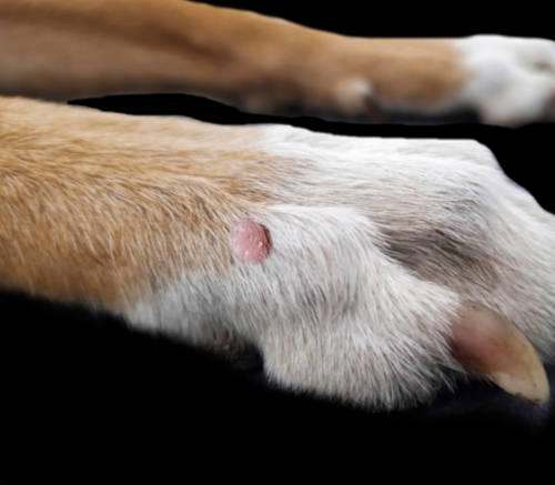
3. Histiocytomas
Histiocytomas are benign skin growths that are relatively common in younger dogs. They are typically small (< 2cm), round, raised, and reddish-pink. Histiocytomas are more common on the head, neck, and ears but can occur on toes and paws, as show on the pictures below.
These growths grow very quickly but are not cancerous, and typically not painful. Most histiocytomas will go away on their own within a couple of months. Larger ones that are bothersome may need to be surgically removed.
Learn more about Histiocytomas or Red Bumps in dogs.
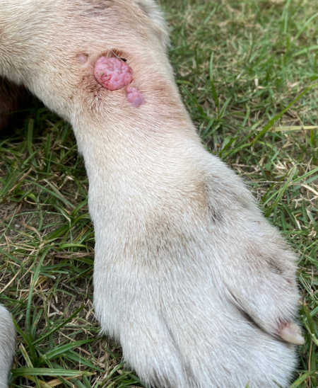
4. Cancerous tumors (three examples of paw tumors)
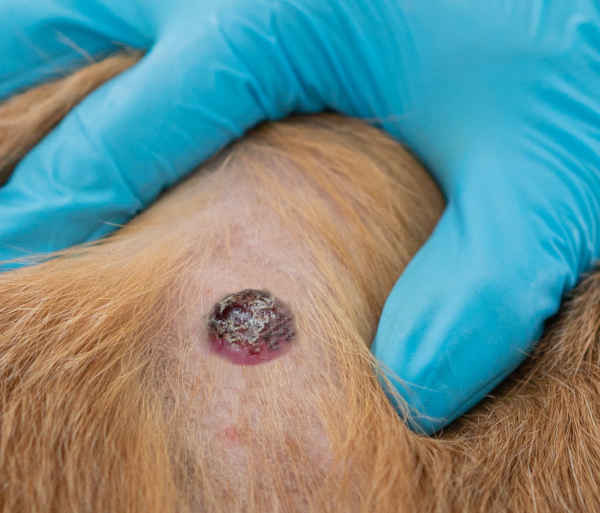
The two main culprits of cancerous tumors or lumps on a dog’s paw are ‘digital squamous cell carcinoma’ and ‘digital melanoma’. Both are typically fast-growing lumps that often change shape over time (see picture below or this picture from VeterianKey.com: squamous cell carcinomas between the toes).
These lumps can ulcerate, bleed, or cause the neighboring tissue to die. Dogs may also have a swollen toe, limp, or lick their foot.
“Cancerous tumors on a dog’s paw can spread to other parts of the body so it’s important to get a diagnosis early.”
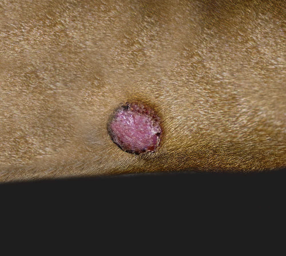
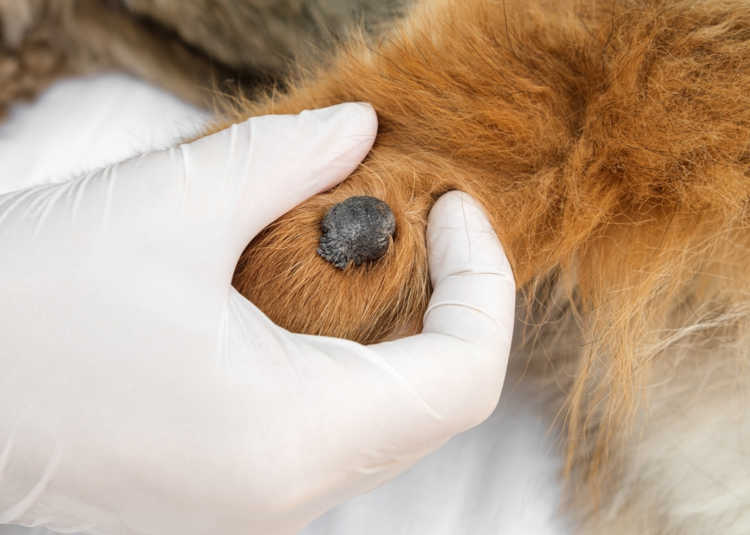
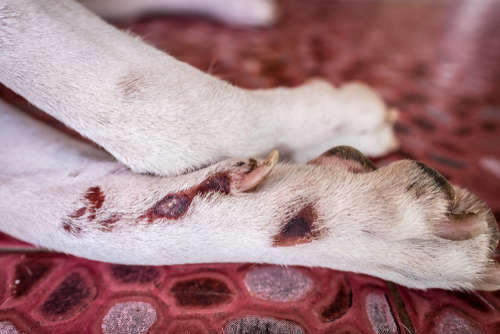
Pictured below is a melanoma on a dog’s leg. Melanomas can occur anywhere in the skin and are the second most common tumor associated with toes in dogs.
Early diagnosis is essential as melanomas that occur on the paws or toes are usually much more aggressive than those affecting other areas. There is a high risk of spread to other tissues and they may damage underlying bone, causing pain and lameness.
If this type of tumor is diagnosed, your vet will often recommend investigations to rule out spread to other organs. Treatment usually involves amputation of the affected toe but may also include radiotherapy and immunotherapy.
Lumps that develop rapidly or cause irritation are more concerning. Monitoring these for an extended period at home is NOT recommended as most malignant skin tumors require surgery, and early diagnosis increases the likelihood of successful treatment.
The prognosis for these paw or toe tumors is very variable depending on the type, size, whether the tumor has spread (metastasis), and what treatment is chosen. If the tumor is removed completely and there is no metastasis, prognosis can be good.
Malignant tumors on a dog’s paw or toes can spread to other parts of the body so it’s important to get a diagnosis early. Learn more about Diagnosing Lumps.
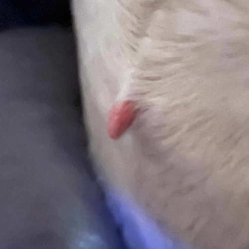
5. Skin tags
Skin tags are benign growths that develop on the surface of your dog’s skin. They come in different sizes, shapes and colors, but are often attached to the skin via a narrow stalk (leaving them dangling in space). Below is a collection of pictures showing skin tags (including a skin tag on a paw).
Learn more about Skin Tags.
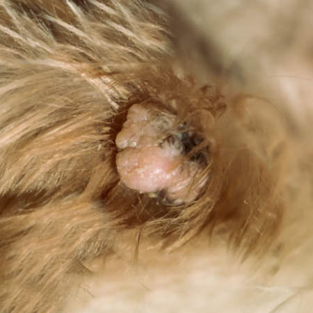
6. Papillomas
Papillomas, also known as warts, are common in dogs. They are caused by a papillomavirus and, in many cases, resolve on their own over months to years. Papillomas usually appear as small raised lumps, which commonly have an irregular, bumpy surface and can be pink, tan, or the same color as the surrounding skin.
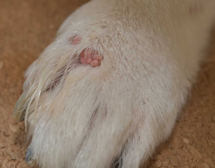
Young dogs are most commonly affected and can have multiple papillomas across the body, especially the face, mouth, and neck.
Papillomas are not typically itchy or painful but, in some cases, can become infected or rarely develop into other types of tumors. If a papilloma is causing irritation, it should be checked by your vet. Papillomas do not typically require treatment, but if they are causing irritation, surgical removal or a biopsy to rule out malignant tumors may be recommended. It is important to regularly check any papillomas for changes, growth, or irritation. Learn more about Papillomas (Warts): Pictures, Treatments.
“It can be challenging to distinguish between a benign lump and a cancerous tumor. A veterinarian’s diagnosis is usually required. Learn more.”
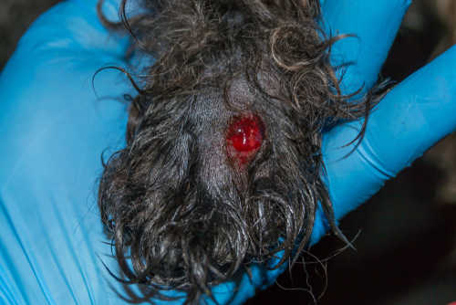
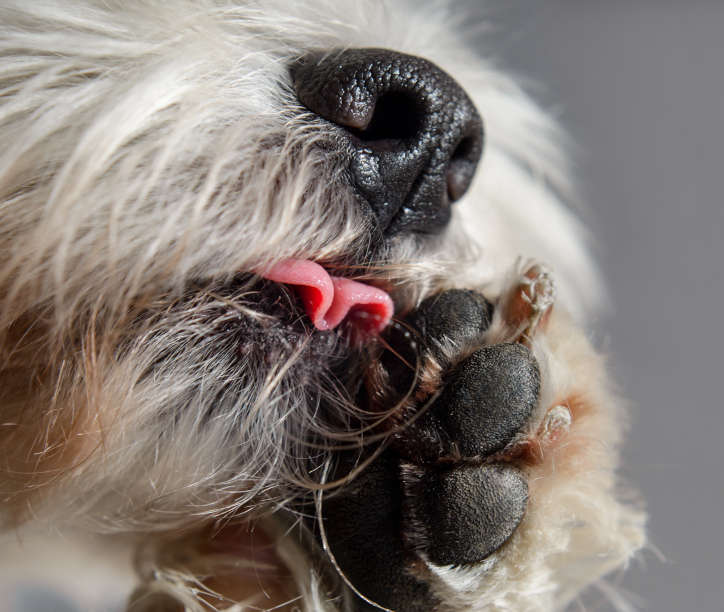
7. Sores due to excessive paw licking (lick granuloma)
A lick granuloma is a lump or lesion brought on by excessive licking. While mild licking can be soothing and even healing for your dog, excessive paw licking will likely be irritating. It may create sores or raised lumps, usually with a raw surface, as shown in the pictures below.
Lick granulomas may bleed or become infected. Many dogs recover well from lick granulomas, although the prognosis is variable depending on the cause and severity of the problem. If lick granulomas are left untreated, they may become more severe and difficult to resolve.
Treatment needs to be aimed at curing whatever is causing the licking. This may be itchiness from allergies or irritants, pain from arthritis, boredom, or anxiety. Once the underlying cause is under control, the wound can be treated with topical or oral antibiotics and an e-collar to prevent further licking (estimated cost: $100-$500 or more for diagnostics and treatment, depending on the underlying cause).
Learn more about Dogs Constantly Licking their Paws.
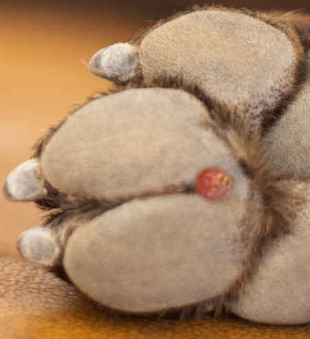
8. Foot Pad Corns
Just like people, dogs can get corns on their feet. These are growths that occur on the foot pad, which is the keratinized part of a dog’s foot. They can be very painful, causing limping and licking of the affected foot. Corns are thought to be caused by a papilloma virus infection, similar to warts, or some kind of repetitive pressure or irritation. View a corn picture here.
Corns are often treated by a veterinarian with ointments, manual abrasion, or surgical removal. Look to spend $100-$500 or more, depending on the treatment of choice. Learn more about Keratoma Corn.
View other types of lumps and bumps:
Pictures of 12 common lumps and bumps on dogs (with vet explanations)
Signs of cancerous lumps include rapid growth, pain, irritation and ulceration of the skin
Generally, lumps that grow quickly or are irregularly shaped and colored indicate a malignant tumor. But not all bad lumps may look like this. Signs that a lump may be cancerous include:
- Rapid growth
- Pain (potentially causing lameness)
- Irritation (causing licking and chewing)
- Ulceration of the skin
- Changes to nail growth or lost nails
Other factors that increase the likelihood of a cancerous lump include old age and certain breed predispositions (e.g. Boxers commonly develop mast cell tumors). Learn more about skin lesions due to cancer.
Diagnosis at the veterinarian: how much will this cost?
Sores & foreign bodies ($50 to $200): as veterinarians, we diagnose many foot-related issues based on a physical examination – lesions caused by infection or foreign bodies are often easily recognized.
In some cases, for example, if a more severe infection is suspected, swabs or skin scrapes may be performed to look for specific bacteria or parasites. This may cost around $50 to $200, depending on the tests being performed.
Lumps & bumps: diagnosis usually involves a “Fine needle aspiration” ($150-$300) or biopsy ($400-$600)
Identifying a lump just by looking at it can be tough. When your vet spots a lump on your dog’s paw, they’ll usually take a sample with a needle to see what cells are inside. Often, the vet can diagnose it by checking the cells under a microscope, but they might also send the sample to a pathologist:
- Simple “fine needle aspirations” (FNAs) cost about $150-300
- Biopsies done under anesthetic may cost $400-600.
- Learn more about Fine Needle Aspirates and Biopsies.
Depending on what your vet initially finds, they may also want to take X-rays. Bloodwork will be done if they suspect cancer to help determine if it has spread.
Take high-quality, clear photos
If you are able to take high-quality, clear photos or video of a lump, your vet may be able to give a reasonable idea of the likely cause via email or video call. It can be helpful to use an object like a ruler or coin for scale if taking close-up photos. For a definitive diagnosis, however, a physical exam & further tests will be needed. In many cases, your vet will need to see your dog in person.
Always get a new lump checked out by your vet
In almost all cases, a new or changing lump on your dog’s toes should be checked by your vet. Many of these lumps are treatable but require veterinary attention (e.g. foreign bodies, infections, interdigital cysts etc.) and may progress and cause pain and complications if left untreated.
Lumps like warts and histiocytomas do not require any treatment, but some malignant tumors may be mistaken for these benign lumps. Delayed diagnosis and treatment of a cancerous tumor can have serious consequences for your dog’s health and prognosis.
Three steps you can take at home to help with paw bumps
Any new lump or bump should always be checked out by your veterinarian. That being said, there are some instances when you can first try at-home treatments. For example, if you know that the cause of a paw lump is a foreign body or an interdigital cyst, you may try the following:
1. Clean and soak with warm water and soap
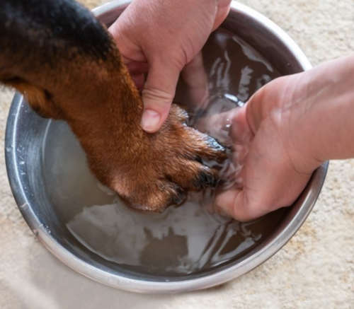
Dog paws can be hard to keep clean. But for minor lumps and bumps like interdigital cysts or foreign body abscesses, that’s just what they need. Clean your dog’s leg with warm water and a mild soap. Rinse thoroughly. You can also soak the paw in warm water and Epsom salts to further cleanse it and to help draw out any little thorns or stickers that may be causing problems.
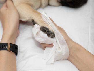
2. Cover it up
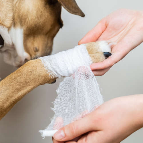
After the paw is clean and dry, you may want to apply a light wrap to continue to keep it clean. This can also keep your dog from working it over with their tongue. A baby or kid’s sock works really well, depending on the size of your dog. Otherwise, a layer or two of vet wrap will do the job. If you’ve never wrapped a dog’s foot before, be sure to get some professional advice first.
Veterinarian Tip: Be very careful when wrapping a dog’s leg or paw! If the bandage is too tight or gets wet and tightens, your dog may lose blood supply and possibly their foot. Bandages on dogs should be placed by a veterinary professional.
3. Monitor it
Lumps and bumps change quickly, so be sure to keep a frequent eye on what’s going on with your dog’s paw. Any changes in size, shape, or color should be immediately seen by a veterinarian. Additionally, if a cyst or abscess doesn’t get better within a couple of days of at-home treatment, see your veterinarian.
Related posts:
 Dog Paw Allergy Issues: 6 Tips to Help Your Dog [With Pictures] - One of my pet parents brought her dog into the clinic because he was furiously licking and chewing on his… [...]
 White Bumps on a Dog’s Paw: Most Likely Reasons - If you’ve noticed a new white bump or lump on your dog’s paw you’re probably wondering what’s causing it. White… [...]
 4 Cysts Often Found on Dog Paws [With Pictures & Vet Advice] - Ahh dog feet, giant Great Dane Marmaduke paws, tiny terrier tootsies, the puffy, fluffy white feet of a Bichon Frisé.… [...]
 Paw Yeast Infections in Dogs: What They Look Like & What To Do - When most people think of yeast, the first thing to come to mind would probably be a freshly baked loaf… [...]
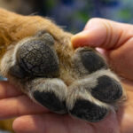 Dog with Cracked Paws? Our Veterinarian Explains What to Do - A dog’s feet often seem indestructible. After all, they’re on the go in all types of terrain, allowing your dog… [...]
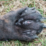 Dog with Swollen Paws [with Pictures] Our Veterinarian Explains What to Do - A dog with swollen paws or paw pads may be suffering from something as simple as a thorn in their… [...]
 Dog Paw Infections and Issues: Pictures & Vet Advice - Dog paw infections are one of the more common issues that we treat in our veterinary hospital: dogs often excessively… [...]
 Why Is My Dog Constantly Licking His Paws? Veterinarian Advice - You may notice your dog licking his paws on occasion. This is often just part of his everyday grooming routine;… [...]
References:
Grassinger, J. M., Floren, A., Müller, T., Cerezo-Echevarria, A., Beitzinger, C., Conrad, D., Törner, K., Staudacher, M., & Aupperle-Lellbach, H. (2021). Digital Lesions in Dogs: A Statistical Breed Analysis of 2912 Cases. Veterinary sciences, 8(7), 136. https://doi.org/10.3390/vetsci8070136
Authors
Disclaimer: This website's content is not a substitute for veterinary care. Always consult with your veterinarian for healthcare decisions. Read More.

My dog has a sore on paw pad, round red bump, it bled badly & had for 15 days, been to vets after it bleeding so badly it would not stop, they bandaged, told no walk, change it every 2 days, bleed is minimal now and on pain relief. Has had 5 vet checkups said continue. My concern is it is not a cut, its a lump like a cyst or corn. Even though bleed is minimal the lump still is so obvious. Surely steroid cream or lump needs to be removed as it will never heal? My dog is a 13yo golden retriever with no other health conditions known. I have pictures before and current.
Hi there Melissa and thanks for your question.
I agree as it has been there for 2 weeks, it is best to have it examined and possibly biopsied, especially as cancers are more common in older dogs.
I’d also want to check for a foreign body under the skin, such as a fox tail or piece of glass
If the lesion does need to be removed, the sooner we can do this the better.
Dr Linda Simon MVB MRCVS
“The information on this website is not a substitute for in-person veterinary care. Always seek advice from your veterinarian if you have concerns about your pet’s medical condition.”