

This article was updated on March 21st, 2024

Discovering a new lump on your dog’s eyes or eyelids can be a cause for concern. If the lump is located near your dog’s eyes, you may feel particularly worried about the impact on their vision. The good news is that most eyelid masses are benign (95%, according to the Animal Eye Institute).
However, these bumps can still cause irritation, corneal damage, and other issues for your dog, and should be addressed by your vet. Here’s what you should know:
9 types of lumps and bumps often found near a dog’s eyes [pictures + vet advice]
Here are some of the most common conditions that can cause a lump around your dog’s eyes or eyelids:
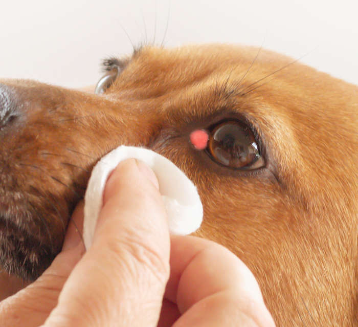
1. Cherry Eye
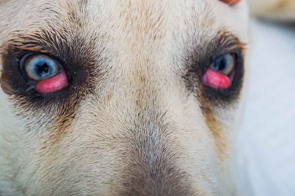
Cherry eye is a prolapse (popping out) of the gland of the third eyelid. It appears as a bulbous red mass at the inner corner of the eye closest to the nose. The condition can vary in severity from a large mass that is consistently present to a smaller mass that comes and goes as the gland pops in & out of place.
In mild cases, cherry eye can be repositioned by simply using a soft cloth and pushing it back in. However, when the glands often pops out, surgery is required in most cases to fix the problem permanently.
2. Meibomian gland tumors
Many eyelid tumors are overgrowth of the meibomian gland, as shown in the picture below.
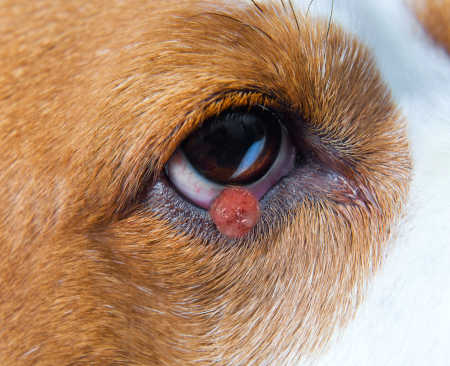
The most common types of meibomian gland tumors are adenomas (which are benign), and adenocarcinomas (which are cancerous). Both tumors arise from the meibomian glands, which line the eyelids and are critical for producing the oily portion of tears.
Benign tumors are the most common and are usually seen in senior dogs. They appear as slow-growing bumps on the inside or outside of the eyelid, and may be pink, pigmented, or lobulated.
Surgery is recommended for large or malignant tumors, and is usually curative. Traditional surgery is effective for removing small masses. If a mass is larger, surgery may be more complex and involve the removal and reconstruction of part of the eyelid.
Learn more about Meibomian Gland Tumors.
3. Papillomas (warts)
Papillomas are bumpy wart-like lesions are typically white, pink, or pigmented, and have a cauliflower-type appearance, as shown in the picture below.
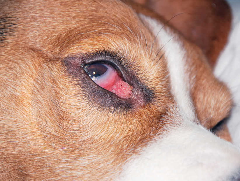
Papillomas are benign and caused by a virus that is most common in younger dogs. Masses may also be seen in the mouth and other parts of the body. Some papillomas will regress without treatment within a few months. Other times your vet may recommend crushing or surgically removing them.
A wart should be removed if it is painful, irritated, growing fast, or if there is suspicion of malignancy.
Learn more about Papillomas/Warts.
4. Melanomas
Arising from pigment-producing melanocytes, melanoma masses either appear as broad and flat or smooth and raised areas on the eyelids. They can also occur in the eye itself. Surgical removal is recommended and generally curative.
Learn more about Melanomas.
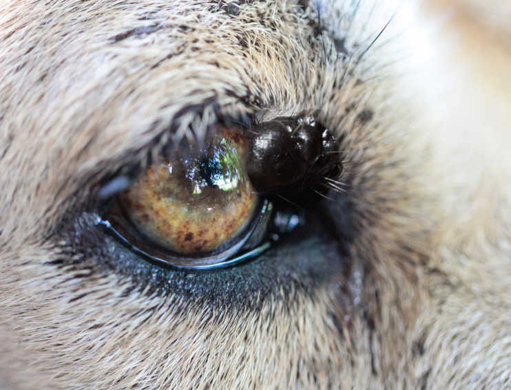
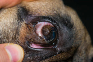
5. Chalazions
You can see a chalazion in the picture below (small dark brown bump on the dog’s eyelid):
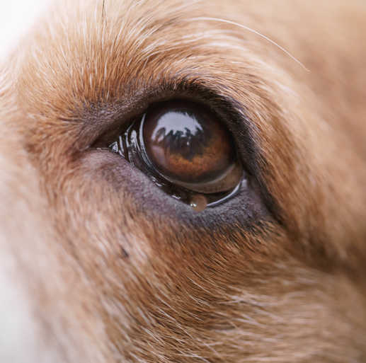
A chalazion is a blocked and inflamed meibomian gland, that appears as a painless, firm, yellow-grey mass on the inner or outer part of the eyelid. They are most common in senior dogs and may be associated with other types of eyelid masses, causing the gland to become blocked.
Whether a chalazion needs to be removed depends on its size, location, impact on the dog’s comfort or vision, and if there’s any sign of infection. Many chalazions resolve on their own over time, but if it persists, grows, or causes discomfort, a veterinarian might recommend an intervention. They may be flushed out under local anesthesia or surgically removed.
6. Sties
A sty (hordeolum) is a blocked gland that has become infected and painful and appears as a red bump on the eyelid. Treatment commonly involves warm compresses to help the sty drain naturally. If there’s an infection, the veterinarian might prescribe antibiotic eye drops or ointment. In severe cases, oral antibiotics may be needed.
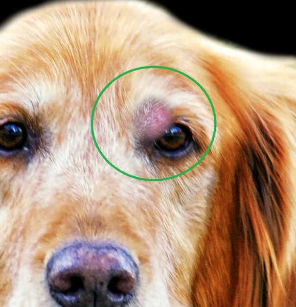
7. Blepharitis, infection, or bug bites
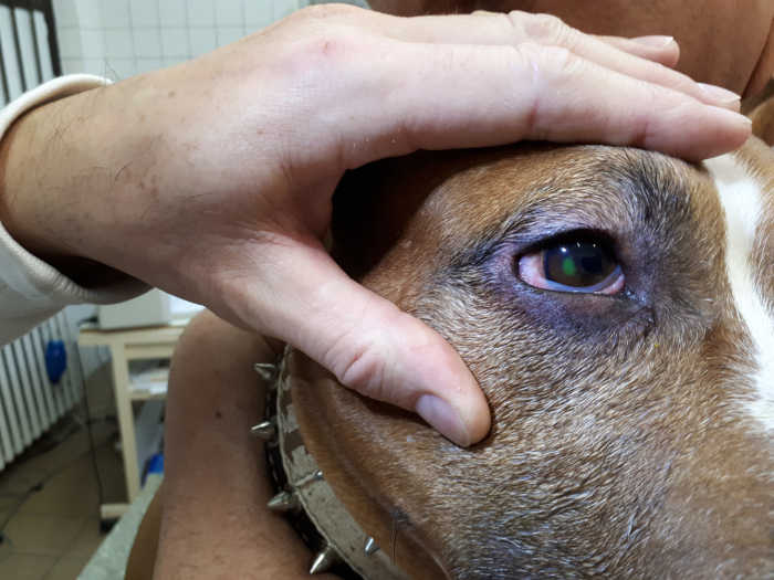
Blepharitis, or inflammation of the eyelids, can occur due to minor bug bites, injuries, masses, infections, allergies, and more. Inflammation generally appears as a raised red swelling that may be tender to the touch, as shown on the picture below:
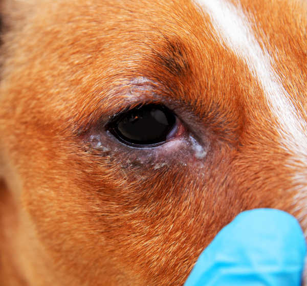
Depending on the underlying cause, it may resolve over time or require veterinary treatment such as antibiotics or anti-inflammatories. Application of a warm compress for 5-15 minutes several times per day may be beneficial.
8. Ticks
Ticks can also attach themselves to the eyelids and have a similar appearance to a mass or skin tag (with the exception of legs), and will need to be carefully removed. Sedation may be needed for this procedure, depending on how wiggly your pup is, to avoid any damage to the eye.
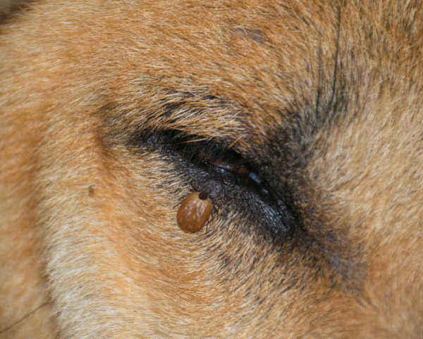
Veterinarian Tip: Owners should never try to remove ticks themselves. A tick that is pulled off will leave its mouth parts in the dog, which will lead to inflammation and infection. Your veterinarian can remove ticks with medications that will cause the tick to release its mouth parts.
9. Other eyelid tumors
Other types of tumors that may affect your dog’s eyelids but are less common include histiocytomas:
as well as squamous cell carcinomas, and mast cell tumors:
Eye or eyelid lumps should be checked by a vet due to the risk of complications
It is best to have any new eye lump evaluated by your veterinarian: masses around the eye can affect tear production or rub on the eye, leading to irritation or corneal ulcers. Additionally, if your veterinarian recommends surgical removal, it is much easier to remove a mass in this area while it is as small as possible. A quick intervention will likely result in a better outcome for your dog.
It is important to see your vet soon if you notice these signs
- A mass that is increasing in size
- A raised or pigmented mass on the eye itself
- A mass that is bothering your dog (causing them to scratch or rub at it)
- Discharge from the eye(s)
- Squinting or excessive blinking
- Color change to your dog’s eye(s)
- A protruding, enlarged, or swollen eye
View more pictures: photo gallery of eyelid bumps
Check out the gallery below or keep reading this article for more pictures and detailed information from our veterinarians:


")


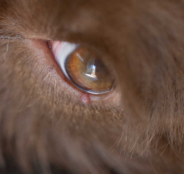
Tumors of the eye itself
1. Melanomas
Tumors can also occur on the surface or the internal structures of the eye, such as the uvea. The most common is melanoma, which typically appears as a flat or raised black/brown area(s) in the eye. Pictured below is an eye melanoma:
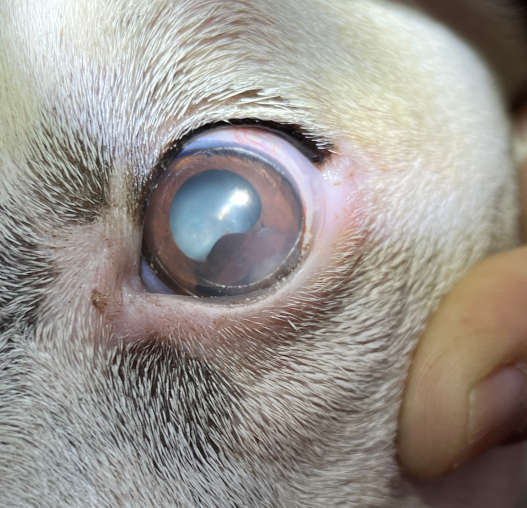
Approximately 20% of uveal melanomas are malignant and have the potential to spread to other parts of the body; however, even benign masses can cause glaucoma (high pressures in the eye) and pain. Removal of the eye is generally the recommended treatment; however, if caught early, specialized surgery may be possible with an ophthalmologist.
2. Dermoids
A dermoid is a benign mass that occurs when normal tissue grows in an abnormal place. While dogs are born with this condition, it may not become evident until later in life. The typical appearance is a hairy growth in the eye. You can view a picture here. As this type of mass can cause irritation, removal is typically recommended and curative.
3. Other
Other types of cancer, such as lymphoma, can also affect the eyes. This can cause a visible mass, swelling, ruptured blood vessels, glaucoma, and pain. This type of cancer has the potential to spread and has a poor prognosis.
What you should know about eye or eyelid lumps
While the majority are benign, lumps and bumps in or around the eyes have the potential to cause several issues. Some conditions, such as cherry eye, can disrupt tear production. Other masses cause damage or irritation by rubbing against the cornea or preventing the eye from fully closing, which can result in corneal ulcers. If a mass is malignant (cancerous), it may spread to other parts of the body and affect your dog’s overall health and life expectancy.
What if the dog’s bump is red? Top causes
You may be concerned if you see a red bump around your dog’s eye. Several common lumps found in dogs may be red or pink in color, including the following. The best way to get a diagnosis is to schedule a visit with your vet:
- Cherry eye
- Bug bites or infections
- Sties
- Meibomian adenomas or adenocarcinomas
- Papillomas
- Histiocytomas [View pictures]
- Mast cell tumors [View pictures]
How a vet can help with your dog’s eye issues
Vet diagnostics
There are several tests that your vet may want to perform to further evaluate your dog’s eyelid mass and the eye itself. In many cases, your vet will not be able to determine the type of mass present just by looking at it, and will need to take a sample for further evaluation.
- Fluorescein stain – This test checks for the presence of a corneal injury or ulcer. A small amount of stain is applied to the eye, which will stick to areas of ulceration and glow under black light.
- Schmear tear test – This test is used to measure tear production. It involves placing the tip of a special filter paper inside the lower eyelid and waiting for 60 seconds.
- Tonometry – This test involves gently touching the surface of the eye with a Tonopen to measure pressures within the eye. Intraocular pressure may be elevated in glaucoma or decreased in uveitis.
- Fine needle aspirate – If a mass is large enough, your vet may be able to collect a small sample of cells with a needle to examine under the microscope.
- Biopsy – This involves the removal of part or all of a mass and submission to a pathologist for evaluation (histopathology).
- Bloodwork, x-rays, and ultrasound – If your vet is concerned about the spread of cancer, they may recommend a full body workup known as staging. This can also be important if surgery is recommended, to make sure your dog is healthy enough for anesthesia.
Vet treatments and surgeries
The treatment recommended by your vet will depend on the underlying issue, which will be determined based on history, physical exam, and diagnostic testing. Some cases may resolve on their own or with medical management; however, most eyelid masses will require surgical removal by your vet or referral to an ophthalmologist.
Traditional surgery is highly effective for removing small masses. If a mass is larger, surgery may be more complex and involve the removal and reconstruction of part of the eyelid. Laser ablation, cryotherapy, radiation, pain medications, antibiotics, lubricating drops, and an E-collar may also be needed in some cases.
Tumors arising from the eye itself, such as uveal melanoma, may require enucleation (removal of the eye), or specialized surgery with an ophthalmologist.
Veterinarian Tip:
“Melanomas can be very aggressive tumors that, if left untreated, can lead to the loss of vision as well as death if they spread to other parts of the body. If you notice a color change in your dog’s eye, consult their veterinarian as soon as possible.”

About surgeries to remove eye or eyelid lumps and bumps
The type, cost, and extent of surgery will vary greatly, depending on the underlying condition. Surgery can range in complexity from small mass removal under sedation and local anesthetic to removal and reconstruction of large portions of the eyelids or even removal of the eye, under general anesthesia. Your dog will be sent home with an E-collar, pain medications, anti-inflammatories, eye drops, and antibiotics if needed. Once the mass is removed, it will be submitted for histopathology to definitively diagnose the type of tumor and determine if any additional follow-up treatment is necessary.
Anatomy of a dog’s eyelid
To understand the types of lumps that can affect a dog’s eyes, it is helpful to first review some basic anatomy of the dog’s eyelids. Dogs have an upper eyelid and a lower eyelid to cover and protect the eye through blinking, and help spread tears to keep the eye lubricated. They also have a third eyelid known as the nictitating membrane. The third eyelid is light pink to white in color and extends from the inner corner of the eye (closest to the nose) horizontally across the eye. It is generally not visible in healthy pets.
There are multiple glands (lacrimal and meibomian) located in the eyelids that produce the components of tears, and nasolacrimal ducts which allow tears to drain.
References
Barnette, C. (2022, June 24). Dog eye stye: Symptoms and treatment options. Great Pet Care. Retrieved December 15, 2022
Cost of pet eye surgery treament. Eye Specialists for Animals. (2021, March 22). Retrieved December 15, 2022
Eye Care for Animals. (n.d.). Dermoids. Eye Care for Animals. Retrieved December 15, 2022, from
Gelatt, K. N. (2022, October). Eye structure and function in dogs – dog owners. Merck Veterinary Manual. Retrieved December 15, 2022
Krob, C., & Haeussler, D. J. (2018, May 29). Canine eyelid masses. ACVO Public. Retrieved December 15, 2022
Ramsey, D. T. (2021). Conditions of the Eyelids and Ocular Adnexa in Dogs and Cats. Lecture at Waltham/OSU Symposium, Small Animal Ophthalmology. Retrieved from VIN.
Stoewen, D., & Pinard, C. (n.d.). Eyelid, conjunctival, and peri-ocular tumors: VCA Animal Hospital. Retrieved December 15, 2022
Veterinary Vision Center. (2020, October 3). Your pet’s eyelid lumps and bumps. Retrieved December 15, 2022
Waltman, S. (2021, April 14). Meibomian gland (eyelid) tumors in dogs. VIN. Retrieved December 15, 2022, from https://veterinarypartner.vin.com/default.aspx?pid=19239&id=10194756
Author
Disclaimer: This website's content is not a substitute for veterinary care. Always consult with your veterinarian for healthcare decisions. Read More.