

This article was updated on October 17th, 2023
In this article, you’ll find a collection of pictures and descriptions of common dog lumps & bumps, carefully curated by our experts: Dr. Spiegel, board-certified dermatologist & leading authority on canine skin issues, and Dr. Whittenburg, our veterinarian director.
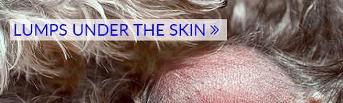
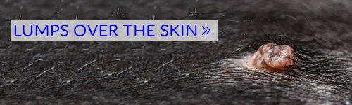
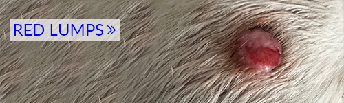
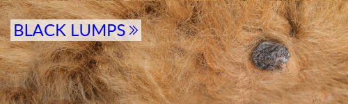
1. Browse by types of lumps
Click on the images below for more information on these types of lumps commonly found on dogs:
2. View a gallery of pictures of common lumps and bumps
Scroll down to view detailed pictures and information about each type of lump, or browse our gallery below to get a quick overview:


3. Twelve common types of lumps and bumps (with pictures and vet advice)
1. Lipomas
Lipomas are the most common benign (ie NON-cancerous) growth seen in dogs. Each lipoma is made up of a group of fat cells that forms a soft round, or oval, lump usually located just below your dog’s skin.
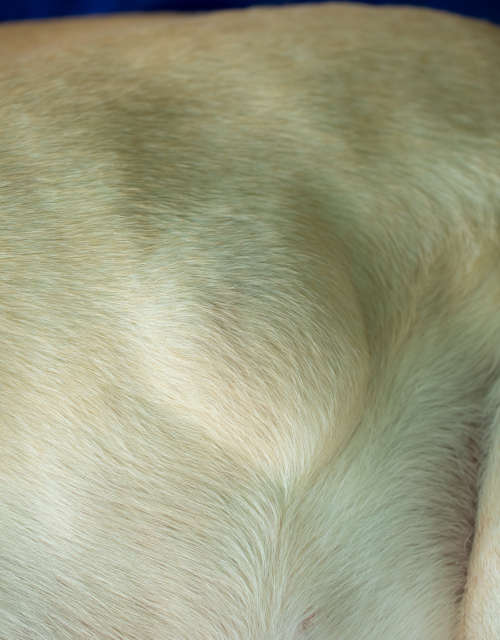
Lipomas are generally easy to move around and don’t feel as though they are connected to deeper tissue in the body. They are usually slow growing and tend to be harmless. These lumps are usually not dangerous, but larger lipomas can cause discomfort or compromise mobility (e.g., infiltrative lipomas).
Treatment: The choice is either to leave them be (benign neglect), or to have them surgically removed. The decision of whether to remove a lipoma or not depends on its location, size, and whether it is affecting your dog negatively.
Learn more about Lipomas.
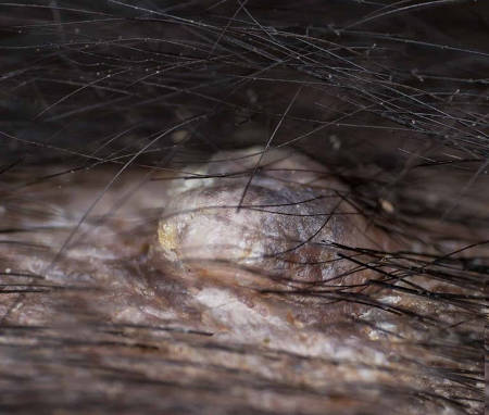
2. Sebaceous Adenomas
Sebaceous adenomas originate from the sebaceous glands. There are the oil-producing glands (sebum) associated with hair follicles and these glands are responsible for maintaining normal skin and hair health and a healthy skin barrier. See pictures below:
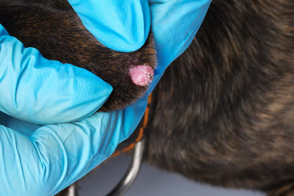
Sebaceous adenomas are usually 4mm to 10mm in size and sometimes extend below the surface of the skin. They tend to grow outward on to the skin surface. These growths are narrower at the base and are often on a thin stalk. They will often ooze a yellow-white oily material.
Treatment: Removal is usually curative, but removal is not usually necessary unless it is getting infected or irritated (sometimes self-mutilation). The prognosis is usually good.
Learn more about Sebaceous Adenomas.
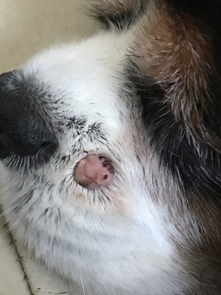
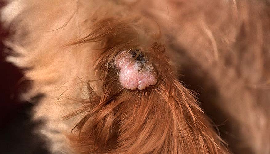
3. Warts
Warts (also called papillomas) are another very common type of old dog lumps. They can show up as a single small lump, or as a group or cluster of tiny lumps which look a little bit like a cauliflower floret. Older dogs tend to develop single warts, whereas puppies are more prone to multiple wart groupings.
Warts on dogs are most often benign and will often disappear of their own accord after a few months. Occasionally they can be, or become, cancerous so any wart that is a long-term issue, or that changes in color/size/look needs to be investigated by a vet.
Treating warts on dogs: Simple lumps like benign warts usually don’t need any treatment unless they get infected, get in the way, or become irritated. This can happen if a dog licks or scratches at the wart, or it is in an area that’s rubbed by a collar or harness.
Learn more about Warts.
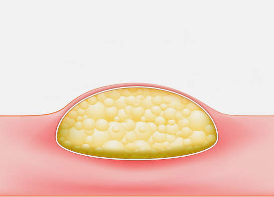
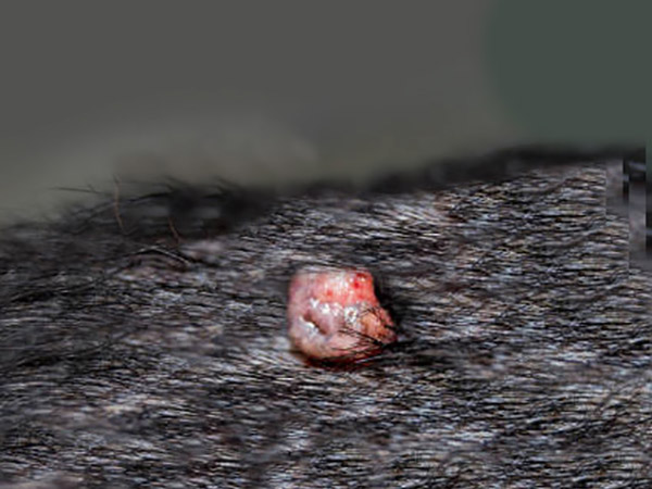
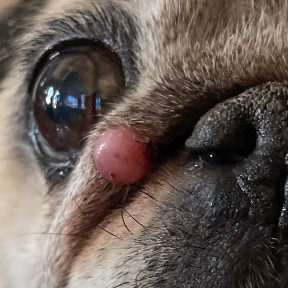
4. Sebaceous Cysts
Sebaceous cysts are common in dogs of all ages and can occur singly, or your dog could have several of them. These types of cysts can be tiny or grow up to an inch or more in size. They tend to resemble a human ‘pimple,’ just larger, as shown on the pictures below:
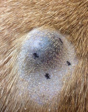
A sebaceous cyst forms when a pore or hair follicle in your dog’s skin becomes blocked or clogged. Matter (oil, skin cells, dirt, and such) collects behind the blocked pore and forms the cyst just below the skin.
Treating sebaceous cysts in dogs: Sebaceous cysts are benign and generally do not need urgent treatment. Some may resolve on their own (‘Come to a head’ and burst or ooze out the pus/gunk inside.) but may “fill-up” again as the cyst lining is still present.
Learn more about Sebaceous Cysts or view 6 Types of Cysts [With Pictures].
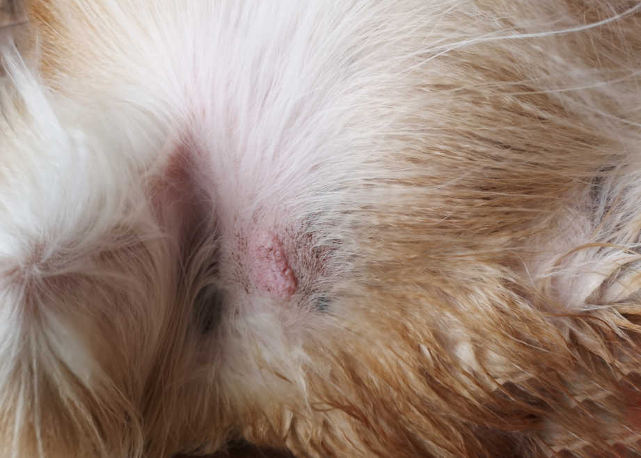
5. Skin Tags
A skin tag is a benign growth that develops on the surface of your dog’s skin. They vary in appearance with a range of different sizes and shapes. Many skin tags are pedunculated – this means that they are attached to the skin via a narrow stalk leaving them dangling in space.
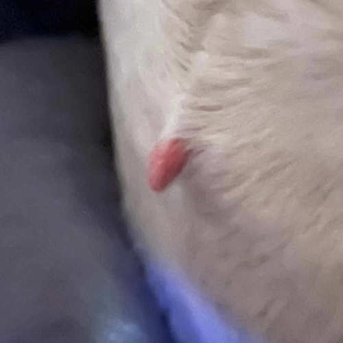
Skin tags are non-cancerous and generally don’t cause any harm to your dog. However, skin tags often develop on areas of your dog’s skin where there is high friction, such as areas that are in contact with the ground or their collar.
Over time, constant rubbing of these skin tags will lead to irritation and may be painful for your dog. In the majority of cases, skin tags are not anything to worry about.
Learn more about Skin Tags.
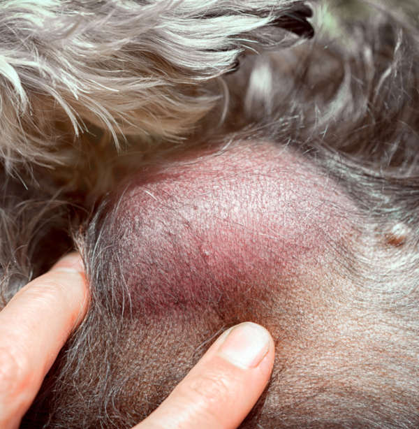
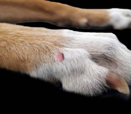
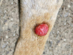
6. Histiocytomas
A histiocytoma is a benign (usually self-resolving transient cancerous) growth found on the skin of young dogs. Common areas affected include the ear flaps, face, feet, and legs. They are dome-shaped and red with no hair (button-like).
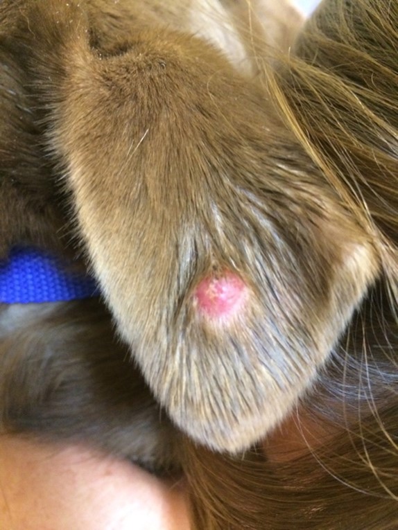
While histiocytomas occur commonly in dogs under three years old, they can affect any dog at any age. Allergic dogs are predisposed.
Most histiocytomas will heal without treatment in 2-3 months. However, in some cases, they can get worse with time. They can look concerning to the untrained eye, and it can be hard to tell them apart from dangerous growths in dogs.
Learn more about Histiocytomas.
Tip from our veterinarians: The lumps seen so far are benign most of the time, but it’s best to biopsy any new lump to confirm. It can be difficult to tell the difference between a benign and a cancerous growth. Needle aspirations and biopsies are easy procedures that will give you peace of mind, and often only require a local anesthetic (lidocaine block).
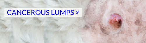
Cancerous Bumps and Lumps
Although most dog lumps and bumps are benign, cancerous tumors can develop. Let’s review the most common types of cancerous lumps in dogs:
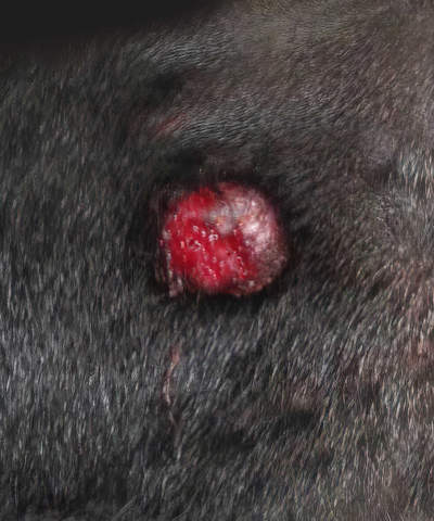
7. Mast Cell Tumors
In healthy dogs, mast cells are the part of the immune system that releases histamine in response to allergies. Mast cell tumors are more often seen in middle-aged and older dogs but can affect younger dogs too. These lumps are very commonly both malignant and aggressive, but it’s impossible to know which ones are and which aren’t without a biopsy.
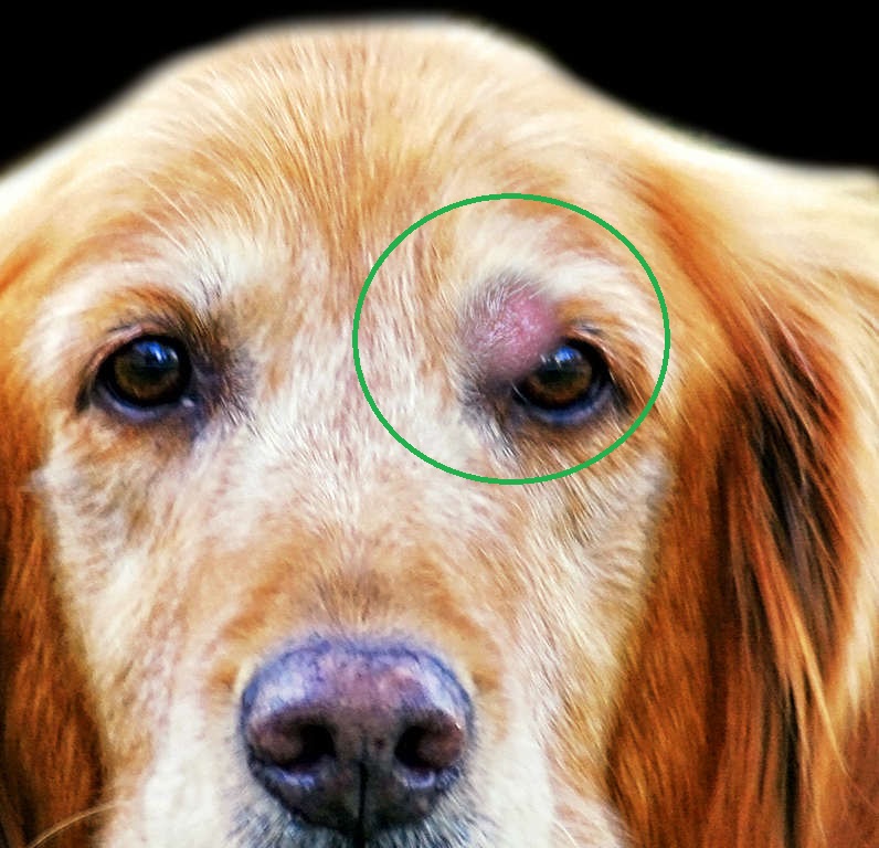
Mast cell tumors can vary in appearance, but usually, they are fairly smooth, round growths visible on the skin surface. Other times they can look like a wart, adenoma or even a skin tag. When they develop beneath the skin surface, they can resemble other masses such as lipomas. Mast cell tumors need to be removed.
Learn more about Mast Cell Tumors.
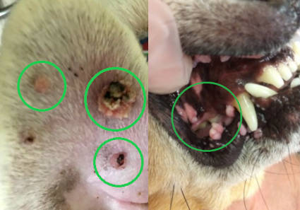
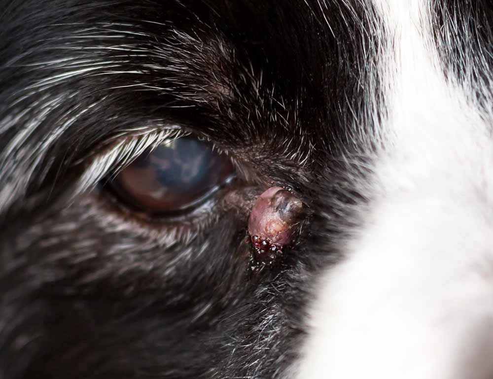
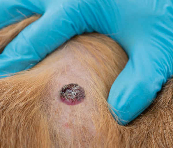
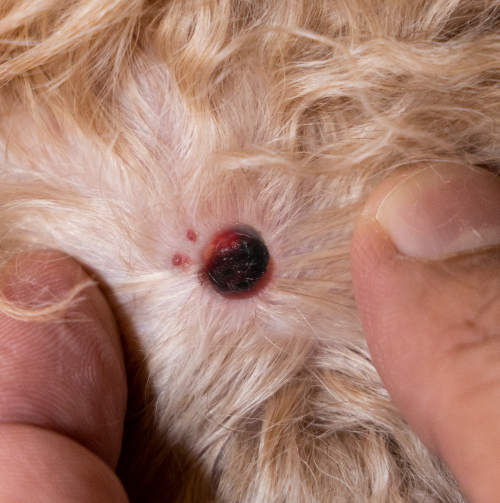
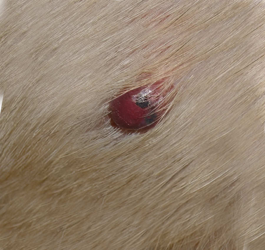
8. Melanomas and Melanocytomas
A melanoma on a dog typically appears as a dark, irregularly shaped growth. They can be small or large, flat or raised. They can appear as a solitary smooth surfaced pigmented nodule, a multilobulated nodule, or even somewhat flat. They can be either benign or malignant, so they shouldn’t be ignored. Below are pictures of a melanoma inside a dog’s mouth and on the lip fold (black growth):

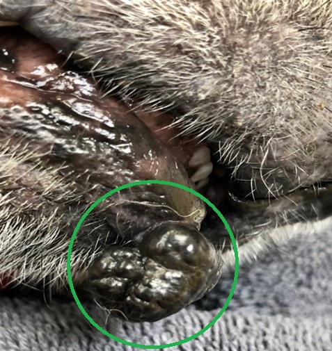
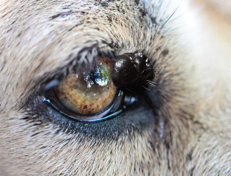
Melanomas/melanocytomas can be detected anywhere, but most malignant melanomas in dogs grow in/around the mouth or in other mucus membranes. When present on mucous membranes, such as around the eyes or lip folds, the prognosis may be less favorable. These growths are also noted on toes (claw beds).
If a dog has malignant melanoma, it’s usually an aggressive cancer that spreads throughout the body quickly, so the lesion needs to be surgically removed as quickly as possible.
Learn more about melanomas.
9. Squamous Cell Carcinoma
Squamous cell carcinomas in dogs are relatively rare, and they are not as aggressive in terms of spreading. However, like some aggressive melanomas or high-grade mast cell tumors, they can also spread (metastasis).
These types of malignant lumps are usually found on areas of skin that are bare, or have little hair, and are more common in dogs with light-colored skin. They can be raised lumps or nodules, or flatter areas of ulcerated skin. They can sometimes resemble adenomas as well as warts.
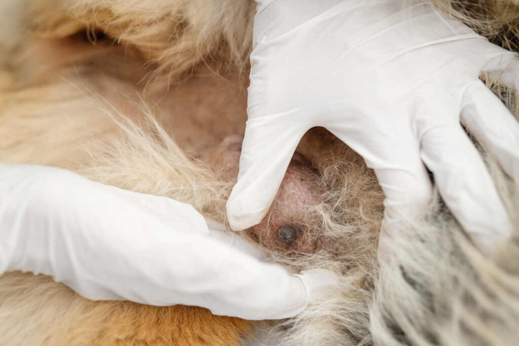
10. Mammary Gland Tumors
Dog mammary lumps come from the mammary tissue along a dog’s belly. Not only are dogs capable of developing breast or mammary cancer, but they do so at an alarming rate, especially if they’re left intact. An intact female has around a 23-34% chance of developing a mammary tumor during her lifetime. Mammary tumors in dogs are benign about half the time and malignant about half the time.
The benign mammary lumps are usually small, firm, and well-defined. You may notice a small lump while petting your dog’s belly, as showcased in our pictures below:
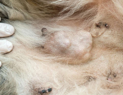
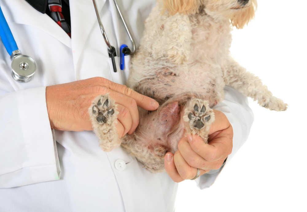
Malignant mammary lumps may be small, large, or a single lump, or multiple lumps. They often have bumpy edges and are tightly fixed to the skin or underlying tissue.
Learn more about Mammary Tumors.
11. Hemangiosarcoma / Hemangioma
Hemangiosarcoma is a malignant cancer of the cells that line the blood vessels of the body. The cells that line the blood vessels start dividing uncontrollably and in an unhealthy manner. This leads to the development of a mass or lump that is very prone to rupture and can even cause a dog to bleed out internally.
Luckily, what is usually seen on the skin are benign blood vessel tumors known as melanocytomas. These are benign blood vessel growths. They may even be in part induced by extended periods of solar exposure.
Learn more about Hemangiosarcoma.
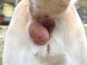
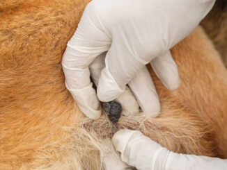
12. Anal Sac Tumors
Anal gland tumors are usually cancerous (The most aggressive ones are adenocarcinomas.) You will notice a growth located next to the dog’s anus, as shown in the picture below.
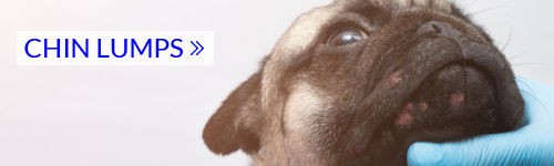
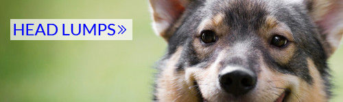
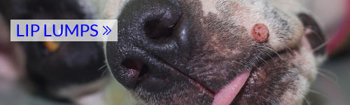
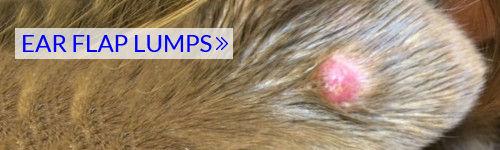
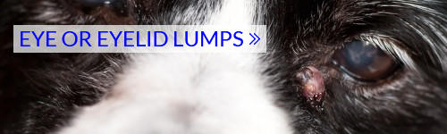
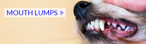
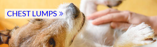
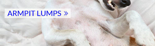
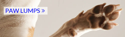
Browse by location
The location of a lump on your dog’s body can sometimes provide clues as to what it is:


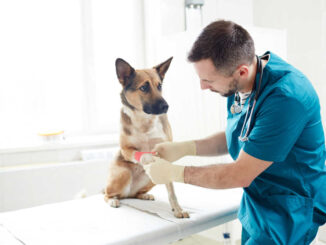
Important: If you find a new lump on your dog, get it biopsied to check for any serious concerns. Needle aspirations or biopsies are easy procedures that will give you peace of mind and ensure your dog gets prompt treatment, if needed.
Diagnosing a lump can cost $150 to $600
Our veterinarian Dr. Linda Simon explains: “Any new lump, bump, or growth needs to be examined by your veterinarian because it is usually not possible to accurately diagnose lumps and bumps from their appearance alone. Cancerous, or malignant, lumps can be small or large. They may itch or cause the senior dog some discomfort. They may do neither.”
Your veterinarian will likely advise sampling the lump and sending the cells to a lab for analysis. The cost of this will vary depending on the clinic and which sort of biopsy procedure is used. Simple FNAs cost about $150-$250, while biopsies done under anesthetic may cost $400-$600. Learn more about FNAs and biopsies.
Not Finding What You Want?
Check out our other pages about skin conditions in dogs:
21 Common Dog Skin Conditions
Skin Lesions and Lumps Due to Cancer
How Do You Know if a Lump or Bump Is Cancerous?
Cancer is the abnormal and rapid growth of previously healthy cells. Therefore, lumps that are growing rapidly or changing in appearance quickly may indicate a more sinister underlying cause. Ulceration, redness, and a firm texture are all common properties of a cancerous lesion, but that doesn’t mean that every lump with these features will be cancerous.
Characteristics of cancerous lumps
You can never say whether a lesion is cancerous just by looking at it, but there are some clues that can help raise suspicions:
1. Rate of growth: Cancerous lesions tend to grow and spread more rapidly than those that are benign. They will grow into the surrounding healthy tissue, causing damage and inflammation. If a lesion doubles in size over the course of a few weeks, then it’s best to get it checked.
2. Texture: Cancerous lesions and lumps are often harder and firmer to the touch.
3. Shape: Due to their rapid and erratic growth, cancerous lumps tend to appear more irregular in shape.
4. Color: Red, black, or just generally ‘unhealthy’ looking lesions may be more malignant in origin.
5. Discharge: Oozing or discharge from the lesion may occur due to damage and death of the tissue in surrounding areas. While any lump can develop a secondary infection, sinister lumps are more prone to producing pus and bleeding.
6. Irritation: A lump that is itchy or causing discomfort to your dog is more likely to be cancerous.
Skin cancer is more common in older dogs also, and a dog’s skin will naturally change with age so sometimes it can be hard to tell whether changes are due to simple old age or due to a more sinister underlying cause. Therefore, it’s important that your vet has a proper look to make the diagnosis – it’s a good idea to have any new lump or bump tested by your veterinarian.
Treating Malignant Skin Lumps
The best way to treat most malignant or cancerous lumps in dogs is to remove them surgically, and as quickly as possible. The smaller the lump the easier the surgery and the less tissue which must be removed. There are also newer injectable treatments for mast cell tumors as well as vaccines to prevent the recurrence of malignant melanoma. Acting fast helps to reduce the chances of the cancerous cells metastasizing (traveling) into other tissues, organs, and lymph nodes. Sometimes radiation therapy, chemotherapy, or other treatments are recommended in addition to removing the tumor.
The exact treatment options will be decided by your veterinarian who will consider the size and location of the tumor and whether it has spread to other areas of the body.
Related posts:
Diagnosing Lumps in Dogs: Fine Needle Aspirates and Biopsies
Cancerous Lumps and Tumors in Dogs: Pictures & Vet Advice
Authors
Disclaimer: This website's content is not a substitute for veterinary care. Always consult with your veterinarian for healthcare decisions. Read More.
I am so grateful for the information it sets me at ease to know what to look for and actually is motivating me to go get my dog’s lumps checked and not procrastinate out of fear