

This article was updated on August 4th, 2023
Tumors in and on dogs’ ears are relatively uncommon, but if you’ve noticed a lump in this area it’s important to get it checked out sooner rather than later; while benign lumps can occur, unfortunately there are a number of cancers that can develop in this location.
To better understand what type of growth you’re dealing with, it’s important to work out where the tumor is. Dogs’ ears consist of 4 parts: the internal ear, middle ear and external ear canal and the pinna. Tumors of the internal and middle ear aren’t visible externally and are most commonly diagnosed once they start to cause neurological symptoms.
The tumors discussed here affect either the external ear canal (visible at the entrance to the ear) or the pinna, which is the cartilaginous fold of haired skin that’s also called the ear flap.
Lumps associated with the ear flap are typically skin tumors, including mast cell tumors, squamous cell carcinoma and histiocytomas. These should be differentiated from hematomas, a common non-cancerous blood-filled type of swelling that typically develops on the outer parts of the pinna as a result of trauma (e.g. headshaking).
Tumors often found in dogs’ ears
1. Ceruminous gland adenocarcinoma
This is the most common type of tumor found in the external ear canal, typically seen in middle aged or older dogs and especially in American Cocker Spaniels. These cancerous tumors can be aggressive, invading local tissues and spreading to other sites including the lymph nodes and lungs.
They usually appear as firm nodules with a broad base, which can be pink, white or mottled with dark pigment in colour. Some will become ulcerated and cause bleeding and irritation. Secondary ear infections may occur resulting in discharge, unpleasant odors and itchiness. Occasionally, tumors that spread to involve the deeper parts of the ear can cause neurological symptoms like a head tilt or circling.
Diagnosis typically requires a biopsy under sedation or anesthetic, and a CT or MRI may be recommended if the tumor is large to identify how far it has spread. Your vet may also recommend investigations to look for tumor cells in the lymph nodes or other areas. Treatment is usually surgical but may be combined with radiotherapy or chemotherapy; with effective treatment dogs can often survive over 2 years. View pictures in this research paper or on PetCoach.co (reader submission) or this picture pre-operative on AnimalCancerSurgeon.com.
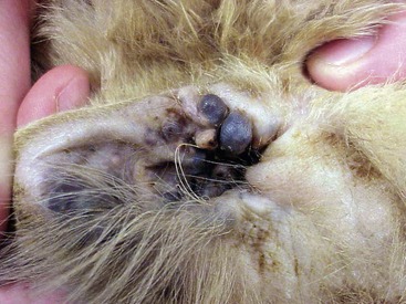
2. Ceruminous gland adenoma
This is the benign version of the tumor described above. Again, they’re typically seen in older dogs and Cocker Spaniels are predisposed. Like adenocarcinomas, they typically grow outwards from the skin of the external ear canal and can be variable in color, shape and size, appearing as nodules or lumpier, multilobulated masses. Occasionally they can also ulcerate, and again secondary infections can occur. It can be very difficult to differentiate these from adenocarcinomas without a biopsy. Although these tumors are benign, surgery may well be recommended if the tumor is large. View a picture of multiple ceruminous gland adenomas in the ear of a cat (On VeterianKey.com).
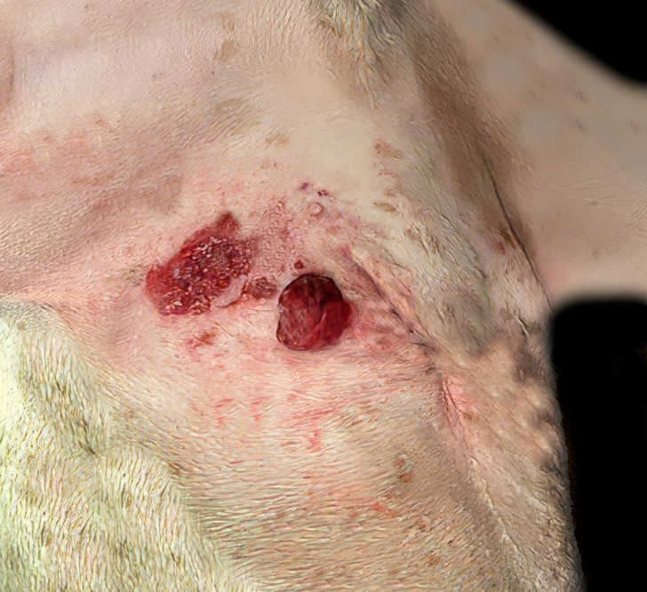
3. Squamous cell carcinoma (SCC)
This type of tumor more rarely affects the external ear canal. It’s a malignant cancer that can have a variable appearance; typically it grows with a broad attachment to the ear, and it often develops an ulcerated, sore surface, sometimes with secondary infection. It’s locally very invasive, so aggressive surgery is the treatment of choice, but radiotherapy can also help.
4. Trichoblastoma (basal cell tumor)
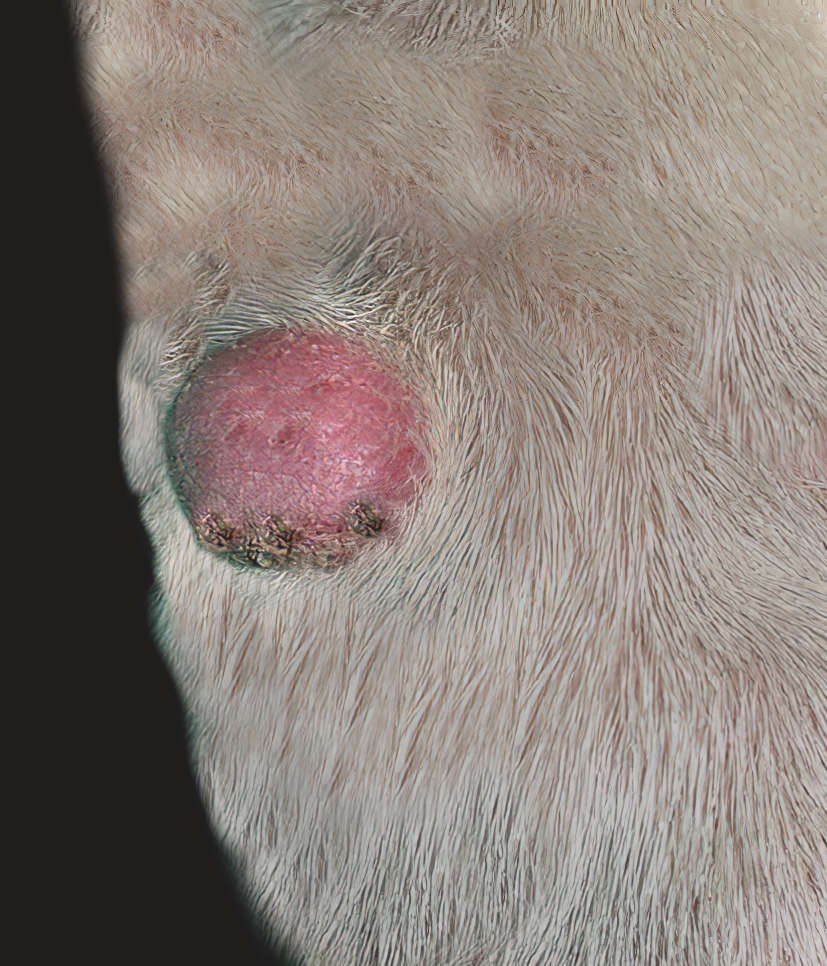
This benign tumor develops from hair follicles and is relatively common at the base of the ear. It usually appears as a single firm dome (or occasionally nodules), with hair loss and potentially ulceration if it becomes particularly large. As a benign tumor, surgery is usually curative but some small tumors may be monitored if they aren’t causing a problem or growing rapidly.
Tumors often found on dogs’ ears
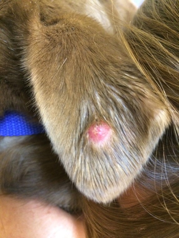
1. Histiocytoma
Histiocytomas are benign skin growths that are relatively common in young dogs, especially on the pinna. They’re usually small (less than 2cm diameter), round, raised and often reddish pink, appearing slightly inflamed. They shouldn’t cause pain or irritation and usually spontaneously resolve after several weeks or months. However, if you notice an inflamed looking tumor, it’s best to discuss it with your vet as it may be worth sampling it to rule out cancer.
2. Mast cell tumor
Mast cell tumors are unfortunately a common skin tumor in older dogs and can occur anywhere, including the pinna. They frequently appear as a pink or red, inflamed, hairless nodule – but these tumors are also known for their highly variable appearance. A fine needle aspirate is usually diagnostic, and treatment typically includes surgery, with further treatment like chemotherapy if there’s evidence of spread to other organs.
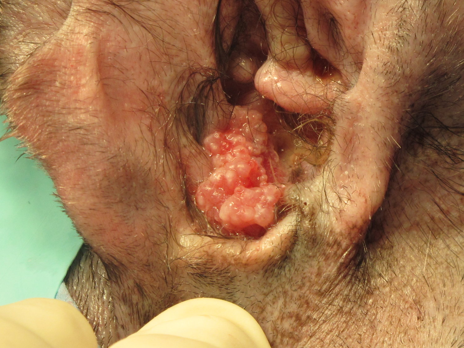
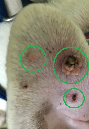
3. Papillomas (warts)
Warts, caused by a papillomavirus, are common in young dogs on their face and neck, and occasionally the ears. These benign growths appear as small, hairless, non-painful cauliflower-like growths from the surface of the skin, and can be variable in color. They’re nothing to worry about – but if you notice a lump, it’s worth checking with your vet to rule out other causes.
What to do if you find a tumor in your dog’s ear
Tumors on dogs’ ears are uncommon, and if you do spot one it’s worth getting it checked by your vet. Unfortunately, many of these tumors are cancerous so will require treatment, but some benign masses are also seen in this area.
It’s important to get a diagnosis sooner rather than later – some of these tumors invade deep into tissues or can spread to other organs so delaying treatment may reduce the chance of a good outcome.
If a tumor is blocking the entrance to the ear canal, it may also increase the likelihood of secondary ear infections, so it’s important to monitor your dog for signs of infection – including head shaking, smelly discharge and scratching at the ear.
Veterinarian diagnosis
Some tumors can be diagnosed based on a fine needle aspirate (a sample of cells taken with a small needle), which can be assessed under the microscope. For others, however, a larger biopsy may be required under sedation or anesthetic in order to get a definitive diagnosis. Your vet may also need to treat any infection that’s present, which may require further tests like culture and sensitivity to identify the bacteria responsible and choose an effective antibiotic.
References
Ear Canal Tumors in Dogs and Cats – University of Pennsylvania Ryan Veterinary Hospital. https://www.vet.upenn.edu/docs/default-source/ryan/oncology-handouts/ear-canal-tumors_ek-ks.pdf?sfvrsn=afb102ba_4
Ear Canal Tumors in Dogs and Cats – University of Pennsylvania Ryan Veterinary Hospital. https://www.vet.upenn.edu/docs/default-source/ryan/oncology-handouts/ear-canal-tumors_ek-ks.pdf?sfvrsn=afb102ba_4
Author
Disclaimer: This website's content is not a substitute for veterinary care. Always consult with your veterinarian for healthcare decisions. Read More.