

This article was updated on January 16th, 2026
Lumps and bumps on dogs’ chests and rib cages are a common finding in our vet clinic, especially in older patients. We frequently see benign masses like lipomas, sebaceous cysts, and adenomas, as well as malignant growths. In this article, we’ll take a look at the common lumps we see on chests and near the rib cages [with pictures].
Benign (non-cancerous) chest lumps
Below, we’ve described lumps and bumps commonly located on a dog’s chest or rib cage, which usually turn out to be benign. Keep in mind, however, that it is usually not possible to tell if a tumor is benign or cancerous just by looking at it. We always recommend consulting with your veterinarian when your dog has a new lump.
1. Lipomas (fatty lumps)
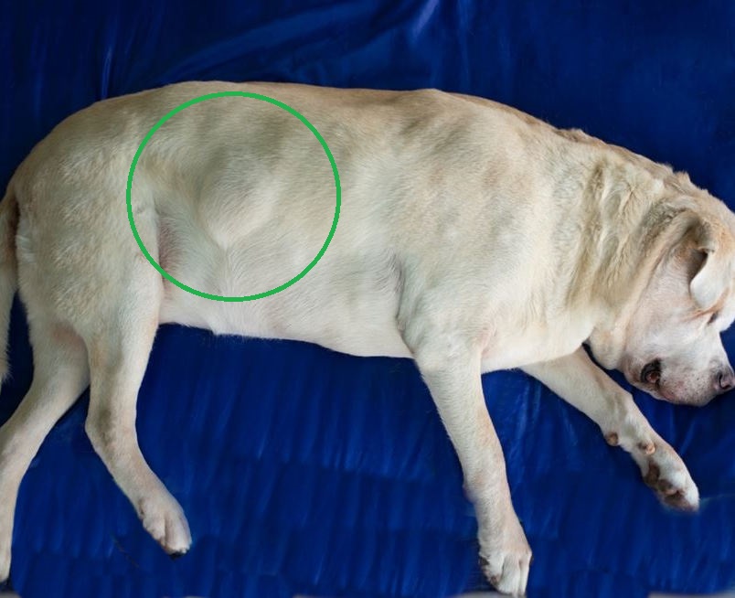
Lipomas are one of the most common lumps I see on dogs’ chests in our practice. These benign (non-cancerous), slow-growing tumors develop from fat cells under the skin. They can be any size and are usually smooth, soft, and non-painful. The overlying skin usually appears normal and moves easily over the lump.
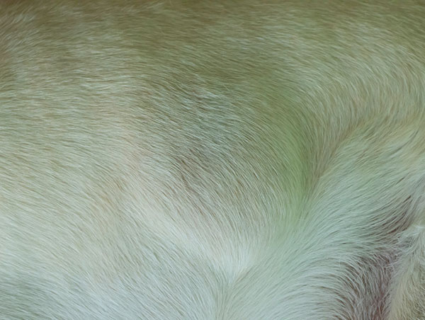
Lipomas are benign, so do not require treatment unless they grow very large or get in your dog’s way. Occasionally, other lumps like mast cell tumors can appear similar to a lipoma, so it’s worth discussing a fine needle aspirate biopsy with your vet to confirm diagnosis. Learn more about lipomas in dogs.
“A large proportion of the older Labradors and Golden Retrievers I see in my clinic will have at least one lipoma. Still, do get any new lump checked over, regardless of what you think it is likely to be.”

2. Sebaceous cysts
Sebaceous cysts are common and develop when a pore or follicle becomes blocked, causing oily secretions to accumulate. They are usually slow-growing, firm, or fluid-filled on palpation and range from a few millimeters to a few centimeters in diameter.
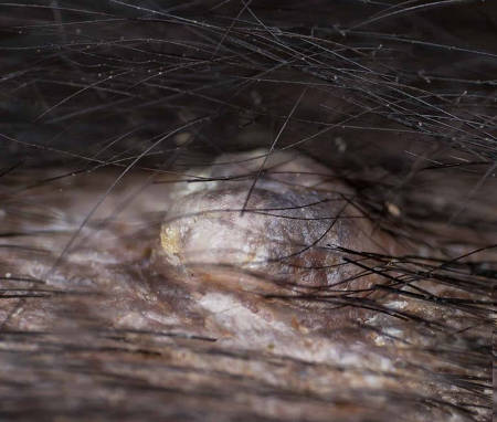
These are often diagnosed based on the appearance of the material within the cyst when a fine needle aspirate is taken. It is important not to assume a new lump is a cyst (even if your dog already has other cysts), as other types of cancerous lumps may be mistaken for a cyst. Most cysts do not require treatment, unless they become infected or inflamed. Learn more about sebaceous cysts.
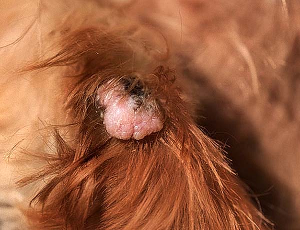
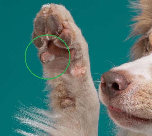
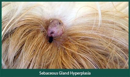
3. Benign sebaceous gland tumors
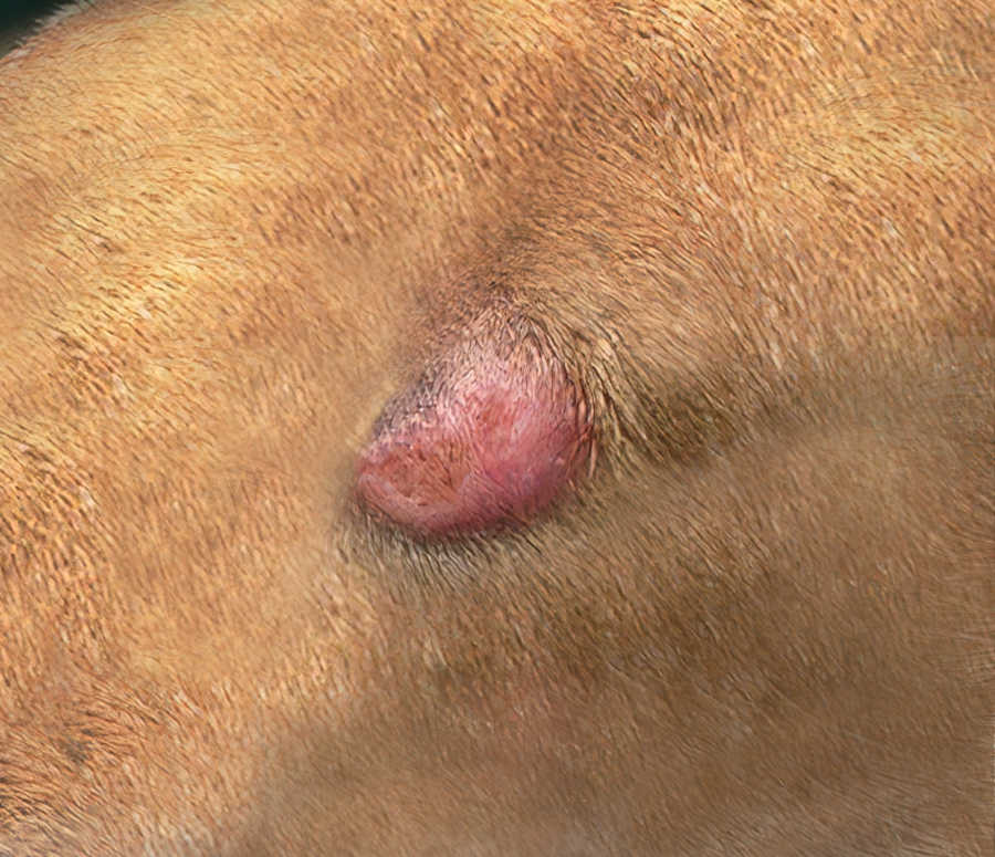
These lumps, including sebaceous adenomas and epitheliomas, are benign tumors associated with sebaceous (secretory) glands. This kind of benign growth is relatively common on the chest of middle-aged to older dogs. Some dogs develop multiple lumps due to a benign condition known as sebaceous hyperplasia.
You can also check this Sebaceous Gland tumor on DermVets.com for another picture example. These tumors often appear similar to warts: raised, hairless, often pale pink but sometimes pigmented small lumps, usually growing slowly and remaining under 2.5 cm in diameter. They can be smooth or have an irregular surface like a wart and occur anywhere on the skin, including the chest.
These benign lumps usually do not require treatment unless they are causing irritation, in which case surgery is usually curative. These are often diagnosed based on clinical examination, but to definitively rule out a malignant tumor (e.g. a sebaceous adenocarcinoma), a fine needle aspirate or biopsy may be recommended. Learn more about sebaceous adenomas.
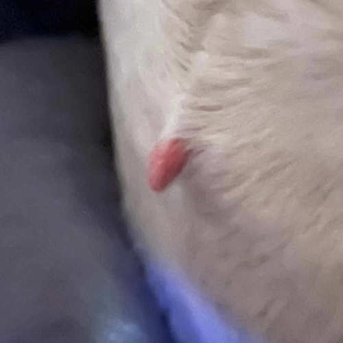
4. Warts and skin tags
Papillomas, also known as warts, are relatively common in young dogs. They are caused by a papilloma virus and typically resolve in several weeks to months. They appear as raised, irregular, ‘cauliflower-like’ small lumps anywhere on the body (including the chest), although they are often seen on the face and neck.
Skin tags are benign growths of the skin that may appear anywhere, at any age, although they are more common in older dogs. They are usually small, soft, and fleshy and do not cause irritation unless they are caught on objects. In the chest area, nipples may occasionally be mistaken for skin tags.
Neither of these lumps typically requires treatment unless they are causing irritation or a wart is failing to resolve; however, if you notice a new or changing lump, it’s still recommended to get this checked by your vet to rule out any more serious diagnoses. View more pictures of dog warts or skin tags.
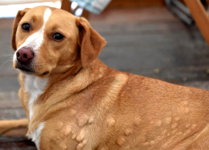
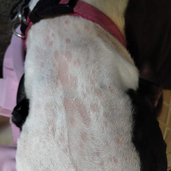
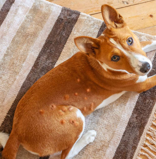
5. Urticaria (hives)
Hives are a not uncommon inflammatory response to a range of possible factors, including insect bites, food allergies, medications, and environmental irritants, including pollen or chemicals. They can occur in any dog and often appear very rapidly, causing raised, itchy, variably sized red bumps or welts across the face and body, including the chest.
In severe cases, swelling of the face, lips, and throat may occur, which can be life-threatening. If you notice swelling in these areas or difficulty breathing, you should seek urgent veterinary attention.
Hives will often resolve over a few hours to a few days, but veterinary treatment can often accelerate this and help prevent complications, especially in more severe cases. If the cause of the hives is identified, avoiding the trigger or allergen is the best option. Home remedies such as oatmeal baths or cold compresses may provide some relief for mild cases. Learn how to treat dog hives at home or view more pictures of hives.
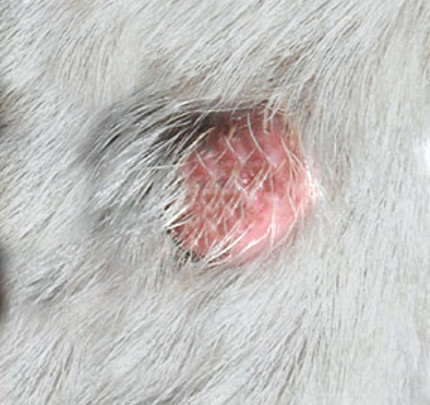
6. Histiocytomas
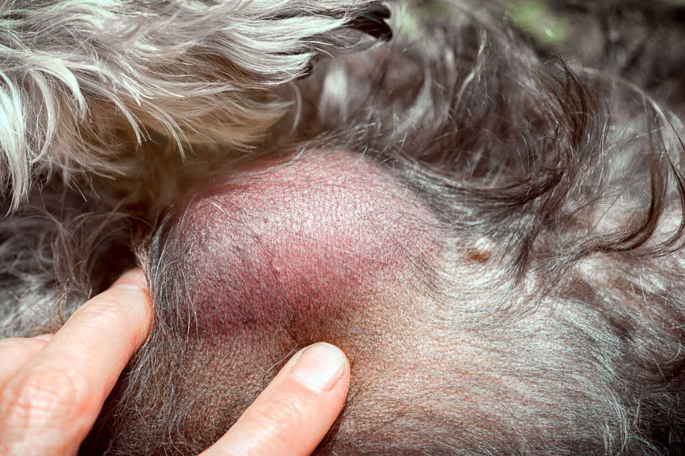
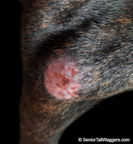
Histiocytomas are a relatively common benign skin tumor typically seen in young dogs. They are usually small (< 2cm), round, raised, pink to red hairless lumps, which do not cause significant pain or irritation despite often appearing inflamed. They are more common on the head, neck, and ears but can occur on the chest area or near the rib cage.
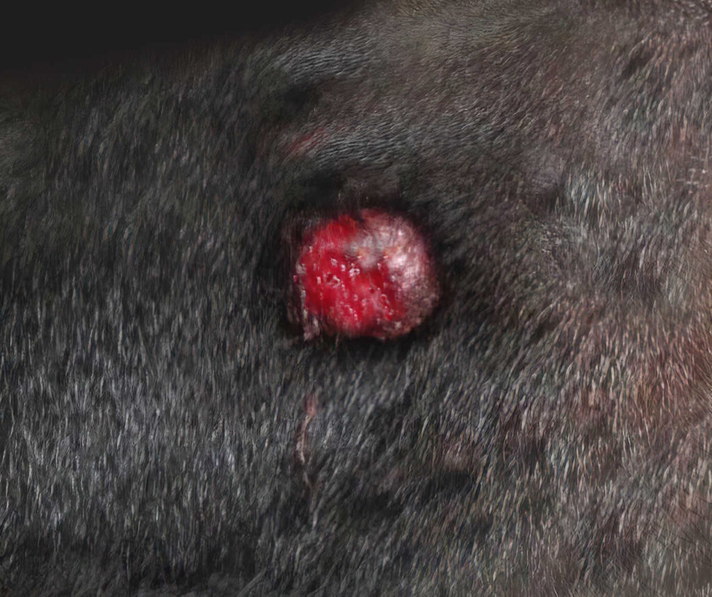
These lumps are benign and regress without treatment in weeks to months; however, they can grow rapidly and appear similar to malignant masses (e.g. mast cell tumors), so testing is often recommended to rule this out. Learn more about Histiocytomas (Pictures & Treatments).
7. Abscesses
Abscesses and swelling due to infection are less common on the rib cage as they usually occur as a result of a wound. Occasionally, small wounds may go unnoticed in this area and allow infection.
Abscesses usually develop rapidly and are painful to your dog. They often appear red and inflamed and may ooze pus or discharge. If any lump appears angry and painful, you should get this checked by your vet as soon as possible, as infections often worsen and spread over time. In the meantime, preventing your dog from licking or scratching the area is essential, and if discharge is present, gentle bathing with dilute salt water may help.
The prognosis for these lumps is usually good. Treatment usually involves cleaning the area, antibiotics, and pain relief. Some cases may require the abscess to be drained or surgically explored under sedation or anesthetic.
8. Injection site reactions
If your dog develops a lump shortly after receiving an injection (e.g. a vaccination) this is usually a benign, mild inflammatory reaction to the injection. These typically occur in the scruff area over your dog’s shoulder blades. The area may feel a little ‘thickened’, or a more defined lump may be present, and this can be a little itchy. Learn more about vaccine lumps / injection site lumps.
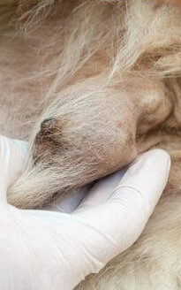
9. Ribs!
It might seem obvious that ribs are found in the rib cage – but in some dogs these ribs can be mistaken for lumps. I’ve seen a number of owners concerned about their puppy or young dog due to a previously unnoticed ‘lump’ on the chest.

In all dogs, the last pair of ribs is described as ‘floating.’ These ribs are short and end partway down the chest, not connecting to the sternum at the bottom of the chest. In some dogs, these last ribs may protrude a little and cause a visible ‘bump’ under the skin.
Malignant (cancerous) soft tissue tumors of the chest
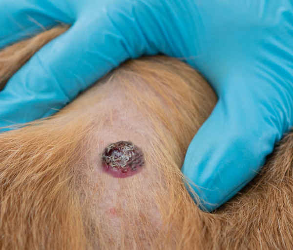
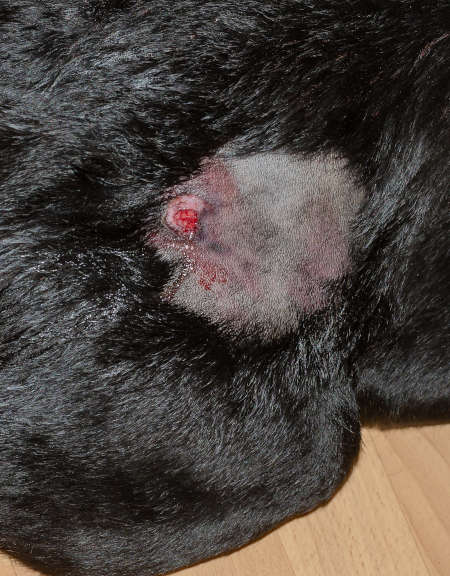
1. Mast cell tumors
Mast cell tumors are a common malignant skin tumor that can occur in any dog, although it is more likely in older dogs and certain breeds (e.g. Boxers). These tumors can develop anywhere in the skin and have a very variable appearance. They often appear as rapidly growing, raised, superficial masses that are often red, inflamed, and itchy, potentially with ulceration of the skin surface.
One sign of a mast cell tumor is a lump that fluctuates in size, as these masses can release inflammatory mediators, which cause temporary swelling. Mast cell tumors can be very serious, and the prognosis for an aggressive ‘high-grade’ mast cell tumor is relatively poor. However, many mast cell tumors are ‘low grade’ or stage 1 mast cell tumors. Surgical removal is usually curative in these cases.
Mast cell tumors are usually easily diagnosed by fine needle aspirate, but in order to grade the tumor, a biopsy is usually required. Treatment usually involves surgical removal, and your vet will often recommend submitting the mass for histology in order to grade it (see how aggressive it is) and determine whether the mass has been fully removed. Your vet may also recommend ‘staging’ to check whether the tumor has spread elsewhere, including local lymph nodes, the spleen, and the liver.
View pictures of Mast Cell Tumors or read our guide to mast cell tumors.
2. Mammary masses
Mammary tumors are very common in intact older female dogs. Spaying at a young age is reportedly protective against developing this tumor later in life, especially if your dog is spayed before her first season. The chain of paired mammary glands in dogs extends from between the back legs up to beneath the chest, and tumors can develop in any of these glands, meaning any lump under your dog’s chest could be related to the mammary glands.
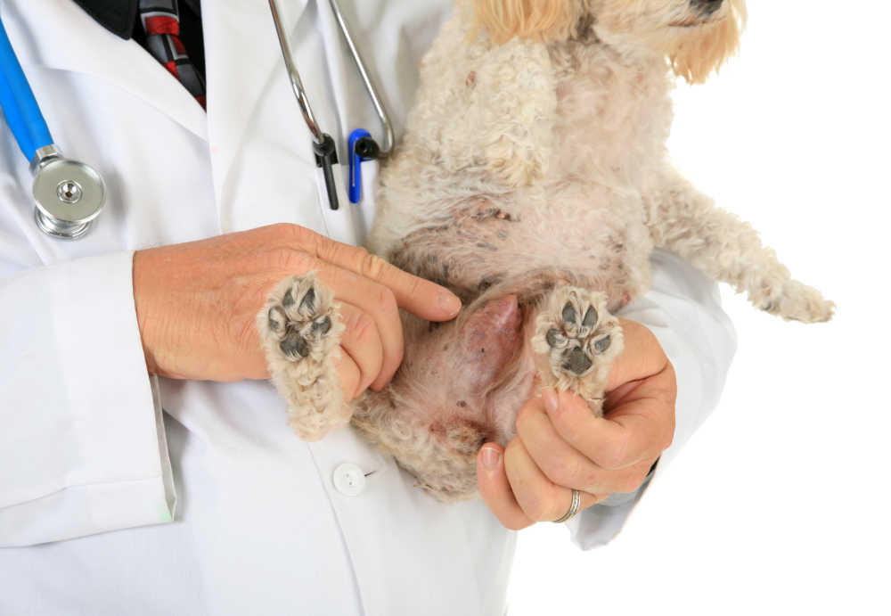
These tumors develop beneath the skin and are associated with the mammary glands, often adhering to the overlying skin. They are usually relatively firm, often irregular in shape, and can be any size. As they progress, some may cause irritation or changes to the overlying skin.
While fine needle aspiration can assist in diagnosing mammary tumors, a larger biopsy is typically required to determine if the tumor is benign or malignant. Additionally, if the mass is malignant, it is important to check for the presence of cancer in other areas, such as other mammary glands, lymph nodes, and lungs.
Typically, surgical removal is the main treatment option, although chemotherapy may also be considered in certain cases. The success rate of the surgery is highest with smaller tumors (less than 1cm), while those larger than 3 cm are less likely to have a positive outcome. Learn more about Mammary Tumors.
3. Soft tissue sarcomas
Soft tissue sarcomas account for around 15% of all skin tumors in dogs. They are a group of malignant tumors, including fibrosarcoma, which usually occur as solitary, slowly growing masses in older dogs. Sarcomas may occur superficially in the skin or deeper beneath it and are typically firm, non-painful, and irregular in shape. These tumors can be seen anywhere on the body, with the chest and trunk being relatively common locations.
Treatment for these tumors consists of surgical removal, and this can be curative. These tumors often grow into the surrounding tissues, however, so local recurrence is relatively common. Due to this, wide surgical margins are often required to fully remove the tumor, which can be difficult is the tumor is large; catching these tumors in the early stages is important. Learn more about soft tissue sarcomas.
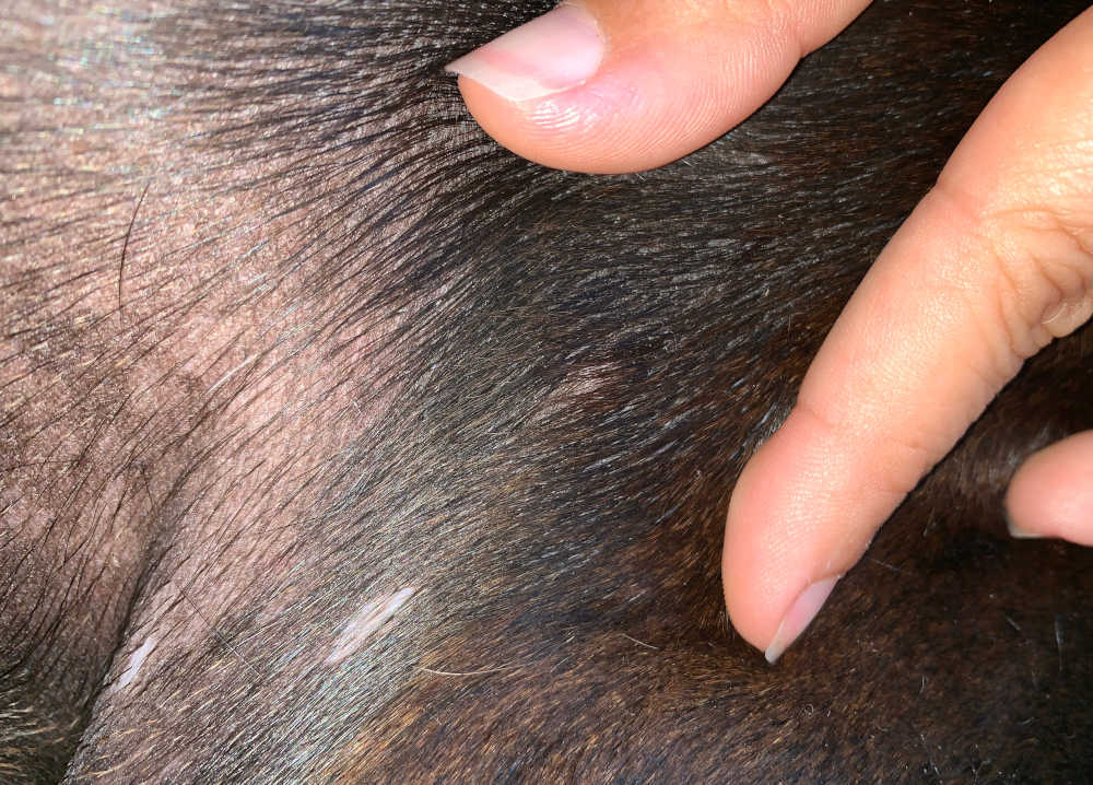
4. Melanomas
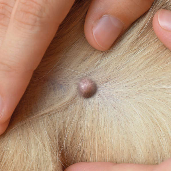
Melanomas can occur anywhere on the skin of any dog, although they are usually seen in older patients. They constitute 7% of malignant tumors in dogs; however, while melanomas on the toes and mouth are frequently highly malignant, those on the skin of the rest of the body are more typically benign.
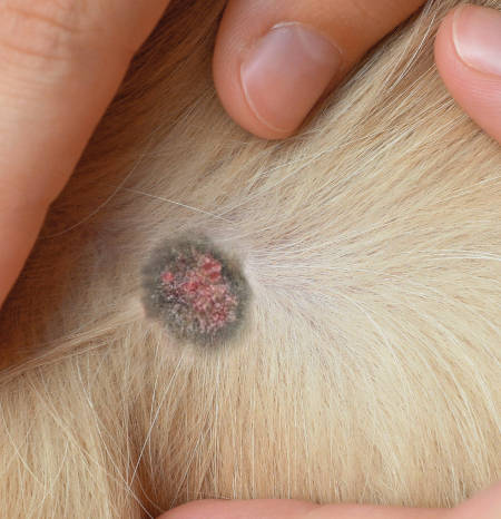
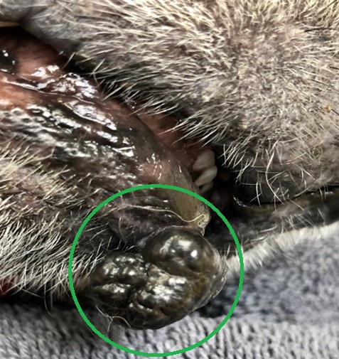
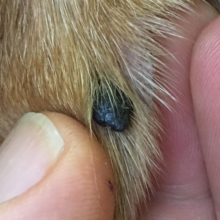
This picture from WalkerVilleVet.com is another good example. On the chest or rib cage, melanomas are usually solitary, raised, round, or irregularly shaped darkly pigmented (black or brown) masses. Occasionally, however, melanomas do not have any pigment. If benign, these masses usually grow slowly.
These tumors may be diagnosed based on a fine needle aspirate, but a more in-depth analysis of a larger biopsy sample (or the whole mass after surgical removal) is usually required to determine whether the mass is benign or malignant definitively.
Treatment for melanomas usually involves surgical removal. If the mass is found to be malignant, further ‘staging’ is usually recommended to rule out metastatic spread to other organs, and if this is identified, further treatments like chemotherapy may be required. Early diagnosis of malignant melanomas is critical for the best possible outcome. Learn more about Melanomas.
Malignant tumors associated with ribs
These tumors are relatively rare in dogs. Tumors affecting the bones are more common in the legs than over the chest area. With these tumors, you would be likely to notice a specific area of immovable hard swelling associated with a rib, which may also be painful. Any lump of this type is likely to require sampling for a definitive diagnosis, but your vet may be reasonably convinced of a presumptive diagnosis based on their clinical examination.
1. Osteosarcoma
Osteosarcoma is the most common type of rib tumor, although tumors of the ribs are relatively uncommon in dogs. These tumors are malignant and metastasize (spread) quickly. They can occur in any dog, although middle-aged to older dogs are more commonly affected.
Treatment options include surgical removal and/or radiation therapy with possible chemotherapy. With treatment, the average survival time of the patient is six months to just under two years.
2. Chondrosarcoma
The second most common tumor of the ribs is chondrosarcoma. This tumor arises from the cartilage of the rib. It is malignant but does not metastasize as often or as quickly as osteosarcoma.
Chondrosarcoma typically occurs in middle-aged to older dogs and can occur in any dog, although some breeds may be more commonly affected.
Chondrosarcoma can be considered cured with complete surgical removal if “clean” (cancer-free) margins are obtained. In areas where complete removal of the cancerous tissue was not possible, the tumor will probably recur, although slowly. Your veterinarian may want to do a second surgery, or depending on your dog’s age, it may be appropriate to monitor the area instead of repeating the surgery.
Frequently asked questions
Are there any symptoms I should look out for if my dog has a new lump?
If the lump is inflamed or painful, oozing any discharge, or your dog seems bothered by it (e.g. licking and chewing) these lumps should be assessed as soon as possible.
Other things to look out for include your dog becoming more lethargic, gastrointestinal signs like vomiting and diarrhea, and drinking more water than usual. It’s also very important to monitor your dog’s weight, as some tumors can cause significant weight loss.
Any of these may indicate a systemic problem like a severe infection, inflammatory reaction, or in some cases, a malignant tumor that has spread to other organs.
Are there any signs I should go back to my vet about a mass we’ve decided to monitor?
If a mass is suspected to be benign or has been sampled and shown to be benign, you may decide with your vet to monitor it. It’s helpful to take regular photos or measurements so you can keep track of any changes. If you notice any of the following, we recommend returning to your vet:
- Sudden rapid growth
- Change in appearance, irregular growth (nodular, uneven as opposed to smooth and round)
- Ulceration
- Oozing or weeping fluid or pus
- Bleeding
- Painful to the touch
When should I see a vet about a new lump?
We recommend seeing your vet about any new lump you notice, as it can be difficult to determine whether they are benign or malignant and whether any treatment is required at home.
If a lump is small, growing very slowly, and causing no irritation, it may be reasonable to wait a short time for a convenient appointment for the lump to be assessed. If you notice a rapidly developing lump, any pain, irritation, or discharge, or any other symptoms like those mentioned above, we recommend seeing your vet as soon as they have an available appointment.
How are lumps and bumps diagnosed?
Not every mass requires a biopsy for diagnosis. There are some lumps that your veterinarian will feel comfortable presumptively diagnosing without a sample. They might treat this lump medically or take a conservative approach and monitor it over time for any changes. The only way to be one hundred percent certain that a lump is benign (not cancerous), however, is for a veterinarian to take a small sample of the lump and have it looked at under a microscope.
For some lumps, a fine needle aspirate is sufficient. This is usually done consciously and involves a small needle (like those used for vaccination) being used to take a tiny sample of cells or other material (e.g. pus from an abscess). For some masses, a definitive diagnosis requires a larger biopsy, which is often taken under sedation or anesthetic.
The cost of an exam for your dog ranges from $50 to $200 nationally, while the average price for a fine needle aspirate is $50-$200. A biopsy is anywhere from $400-$800. Learn more.
Does it tell you anything about the lump if it is hard or soft?
Generally, harder lumps are more concerning than soft lumps beneath the skin. This is mainly due to the fact that benign lipomas (one of the most common types of lump found under the skin) are soft; the fact that a lump under the skin is hard rules out the most common benign diagnosis. There are still other types of benign lumps that are firmer, so this isn’t a reason to panic, but it is a good reason to get a lump checked.
Soft lumps are more likely to be benign lipoma, but this is still not definite – some lumps, like mast cell tumors, can be very soft on palpation. This is why we recommend keeping a close eye on any lump and discussing with your vet further testing for any new lump.
Similarly, some lumps are more firmly attached to underlying tissues while others move around easily, either under the skin (like a lipoma) or with the skin if they have grown from it. Any lump that feels firmly fixed in position is more concerning, as malignant lumps are more likely than others to grow into surrounding tissues.
References
Fonseca-Alves, C.E. et al. (2021) “Current status of canine melanoma diagnosis and therapy: Report from a colloquium on canine melanoma organized by Abrovet (Brazilian Association of Veterinary Oncology),” Frontiers in Veterinary Science, 8. Available at: https://doi.org/10.3389/fvets.2021.707025.
Jackson, H. and Marsella, R. (eds) (2012) BSAVA Manual of Canine and Feline Dermatology. 3rd edn. British Small Animal Veterinary Association.
Stefanello, D. et al. (2008) “Marginal excision of low-grade spindle cell sarcoma of canine extremities: 35 dogs (19962006),” Veterinary Surgery, 37(5), pp. 461–465. Available at: https://doi.org/10.1111/j.1532-950x.2008.00408.x.
Author
Disclaimer: This website's content is not a substitute for veterinary care. Always consult with your veterinarian for healthcare decisions. Read More.

