
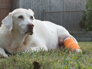
This article was updated on May 1st, 2023
According to research, histiocytomas make up 15.9% of tumors found in dogs, while mast cell tumors make up 17.8%. In our veterinary clinic, we do see mast cell tumors more frequently than histiocytomas. However, these two skin conditions can be very similar in appearance. They can even look identical to each other.
In this article, we’ll look at 10+ pictures and explain the differences and similarities.
1. What is a mast cell tumor in dogs?
Mast cell tumors are the most common skin tumor found in dogs. These tumors form when the immune cells called mast cells, over-replicate. These tumors are often cancerous, and they can spread through your dog’s skin to internal organs.
What do mast cell tumors look like on a dog?
These tumors are known for their unpredictable appearance and the ability to mimic harmless skin conditions. They can look like a large or small bump that is also:
- Red
- Ulcerated
- Flesh colored
- Pink
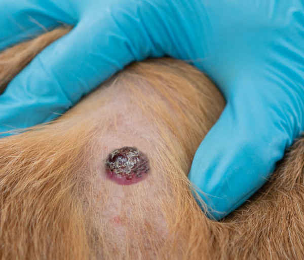
Below is a gallery of pictures showing mast cell tumors that could be mistaken for histiocytomas. Keep in mind that it’s usually not possibly to tell the nature of a lump or bump just by looking at it. These pictures are meant to be for educational purposes only (learn more).
View more pictures of mast cell tumors.
If your dog has a mast cell tumor, they may also have:
- Itching
- Redness
- Vomiting
- Diarrhea
- Lethargy
Can a mast cell tumor be misdiagnosed?
Yes, a mast cell tumor can be diagnosed incorrectly. In fact, according to research, mast cell tumors look similar to the following skin conditions in dogs:
- Histiocytoma
- Lipoma
- Granuloma
- Nodular T cell- lymphoma
Often, an owner will assume a mast cell tumor is a harmless condition like a lipoma (fatty tumor), and they won’t visit the vet to biopsy it. Since mast cell tumors are known to mimic other skin conditions in dogs, new lumps and bumps should always undergo biopsy by your veterinarian.
How quickly do mast cell tumors grow?
They can grow rapidly or slowly. It’s not uncommon for a dog to have a lump for years that grows slowly and out of nowhere, grows large overnight.
What is the best treatment?
The prognosis and treatment depend on the grade, stage, and location of the tumor. Treatment consists of one or more of the following:
- Surgery
- Medication
- Radiation
- Chemo-therapy
- Stelfonta injection
According to research, the best treatment for a mast cell tumor is surgical removal and a combination of other medication and therapies. Mast cell tumors are often cancerous and need to be removed as soon as possible. Even small tumors have the potential to spread to the organs. For more information on treatment and life expectancy, read our article here: Stages of Mast Cell Tumors & Life Expectancy.
2. What are Histiocytomas?
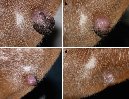
A histiocytoma is a non-cancerous tumor found in dogs under three years old. Histiocytomas form when immune cells called Langerhans cells over-react and replicate rapidly. Treatment is simple. In most dogs, histiocytomas regress (shrink) in 3 months. In rare cases, a dog may have multiple histiocytomas. Multiple histiocytomas are a symptom of a dangerous skin disease called histiocytosis. In histiocytosis, affected dogs have multiple lumps on their body. These dogs also develop granulomas in the bones, causing pain, inflammation, and bone fracture.
What do histiocytomas look like on a dog?
Histiocytomas are small benign lumps that appear most commonly in young dogs. They do not cause itching or pain, and they usually appear:
- Reddish-pink
- Small, less than 2 cm.
- Hairless
3. How to tell the difference between a mast cell tumor and a histiocytoma
It is impossible to diagnose your dog’s lump based on its appearance. However, it is helpful to have a general idea of how different tumors look and behave. Let’s take a look:
Differences in size
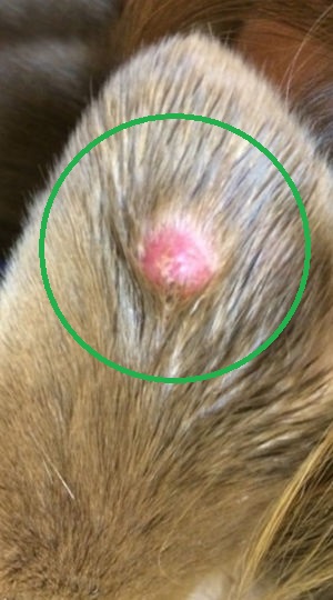
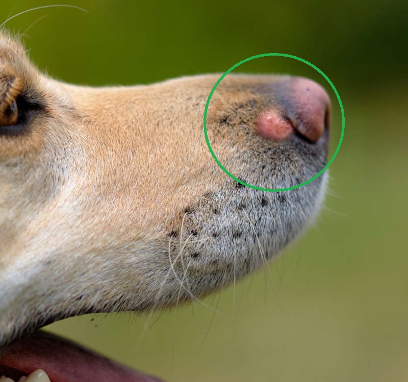
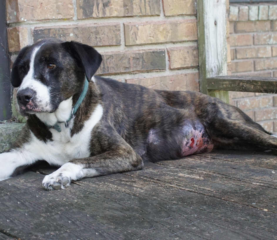
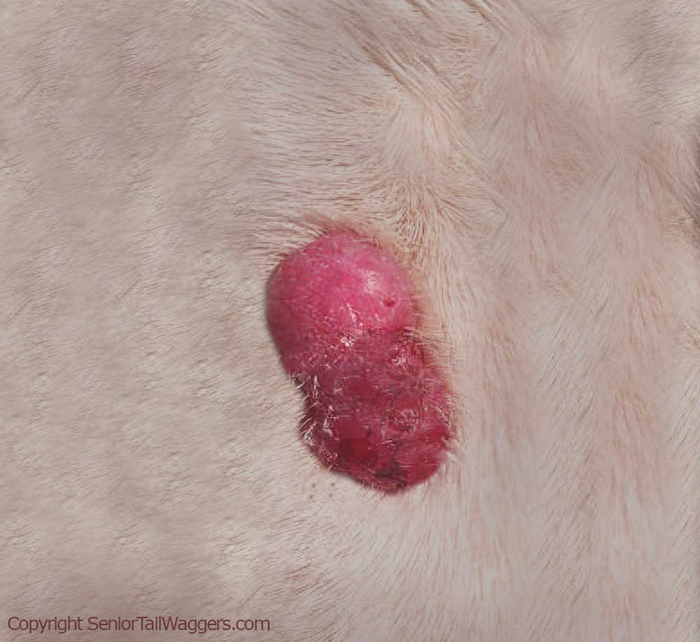
The difference in size: mast cell tumors can be large or small. Histiocytomas are small (usually less than 2cm), but they can be larger. The picture below shows a large mast cell tumor:
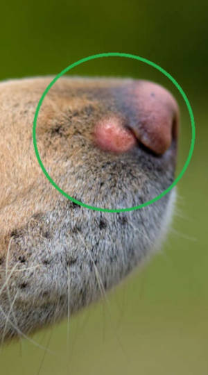
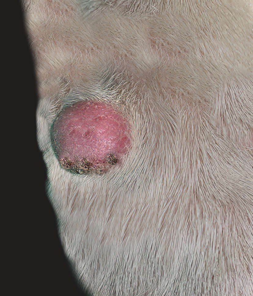
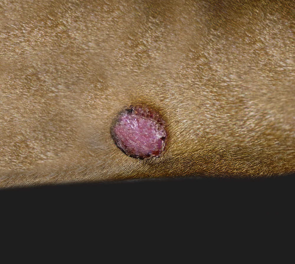
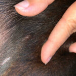
The picture below shows a histiocytoma on a dog’s leg. Most histiocytomas are small and do not get larger than 2cm:
Difference in color
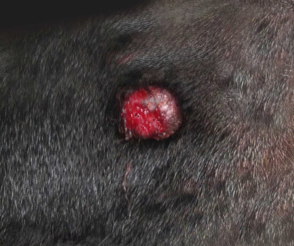
A mast cell tumor can be any color. They are often red, pink, flesh-colored, or even black when they are ulcerated. A histiocytoma is almost always a reddish-pink color. However, they can be black or brown when they are scabbed over.
Difference in feeling
The feel of a mast cell tumor varies. Some tumors are fixed and connected to tissues under the skin, and some are superficial. These tumors can be soft if they are superficial. However, they are usually much firmer.
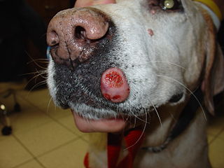
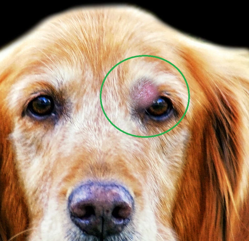
How does a histiocytoma feel? Histiocytomas, on the other hand, are usually soft and moveable. This is a large histiocytoma. Note the soft appearance. Histiocytomas are usually softer than mast cell tumors:
Differences in rate of growth
Mast cell tumors can grow slowly or quickly. It is not uncommon for a mast cell tumor to grow slowly for years and suddenly grow much larger overnight. Histiocytomas usually grow over a 1-4 week period of time.
A helpful way to tell the difference between the two is to pay attention to how long a lump has been present. For example, most histiocytomas will shrink within 3 months. And mast cell tumors tend to grow larger and smaller from day to day, never disappearing completely. Mast cell tumors will wax and wane (get larger and smaller, overtime). However, unlike histiocytomas, mast cell tumors will never go away on their own.
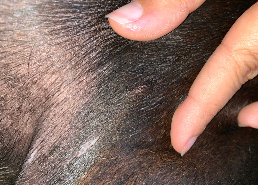
Histiocytoma regression (shrinking):
You will find below a link to a picture showing a histiocytoma slowly disappearing (regressing). As you can see, it looks very similar to the mast cell tumors in the previous photo. However, with histiocytomas, they begin to shrink over time: View picture from VeterianKey.com.
Diagnosing mast cell tumors vs. histiocytoma
Histiocytomas are typically tested the same way as mast cell tumors. First, your veterinarian will do a thorough physical exam and discuss the behavior of the lump. Your vet will want to know things like:
- How long has it been since you noticed the lump?
- Is the lump itchy or painful?
- Does the lump shrink, grow, or change?
- Does your dog have any other symptoms?
Your vet will recommend a fine needle aspirate or biopsy. A fine needle aspirate is a procedure where your vet uses a small needle to collect cells from the lump. After the cells are collected, the vet will look at them or send them out for a pathologist to examine them. Because fine needle aspirates do not always show cancer cells, a surgical biopsy is the best test to diagnose a tumor. Surgical biopsies are collected when your dog is under anesthesia. Your vet will cut out the lump and send it to the pathologist.
Learn more: diagnosing a lump with a fine needle aspirate or biopsy
“I may be a vet who has diagnosed hundreds of cancerous lumps, but I’m still going to sample a lump if I have any degree of uncertainty. Mast cell tumors can take on a range of appearances, so sending the cells to the lab for analysis is usually the most sensible way to reach a diagnosis.”

Related Articles:
 13 Pictures of Mast Cell Tumors in Dogs [With Vet Comments] - Mast cell tumors are the second most common cancerous tumor seen in dogs; they account… [...]
 The Stages of Mast Cell Tumors and Life Expectancy [With Pictures] - When you find a new lump on your dog, it’s easy to dismiss it as… [...]
 Mast Cell Tumors In Dogs: A Veterinarian’s Guide for Owners - Mast cell tumors are one of the most common forms of cancer in dogs. They… [...] What is My Dog’s Life Expectancy With Mast Cell Tumor? A Vet Weighs In - Just this past week, I cared for a dog who was recovering from a large… [...] What to Do When a Dog’s Mast Cell Tumor Itches, Bleeds, or Bursts - Mast cell tumors are a common skin cancer in dogs, accounting for 20% of all… [...]
 When to Euthanize Your Dog with Mast Cell Tumors [Veterinarian Advice] - The worst part about owning a dog is that their lives are far too short.… [...]
Author
Disclaimer: This website's content is not a substitute for veterinary care. Always consult with your veterinarian for healthcare decisions. Read More.

