

This article was updated on October 3rd, 2023

Featured below is our collection of pictures featuring common tumors, cysts, and growths in dogs. This collection has been compiled by our veterinarians with the hope that it will help pet owners and their veterinarians recognize health issues quickly and take prompt action to help their pets.
Pictures of common cysts, tumors, or growths in dogs
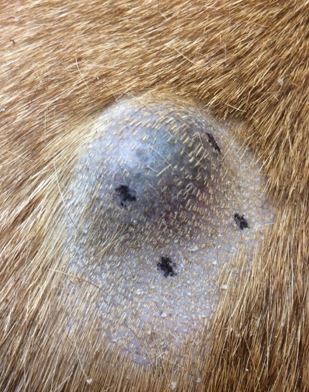
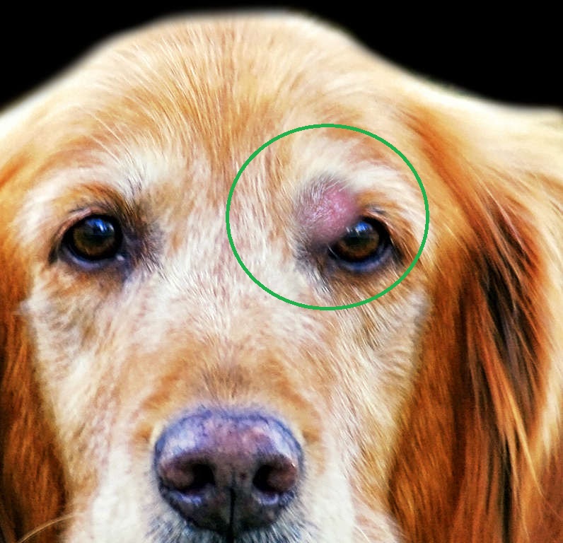
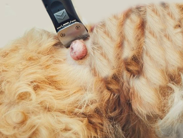
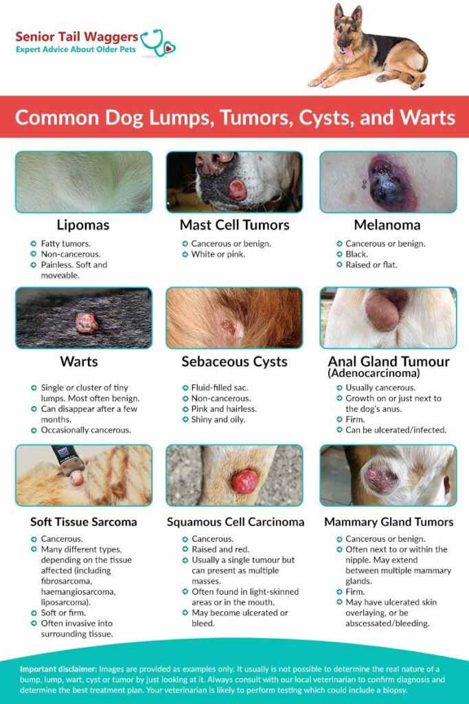
1. Lipomas
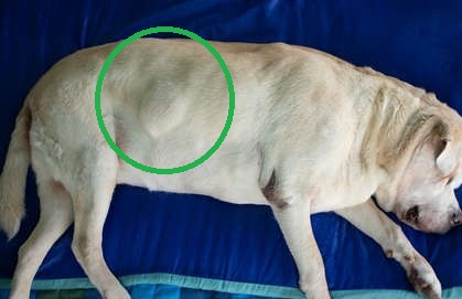
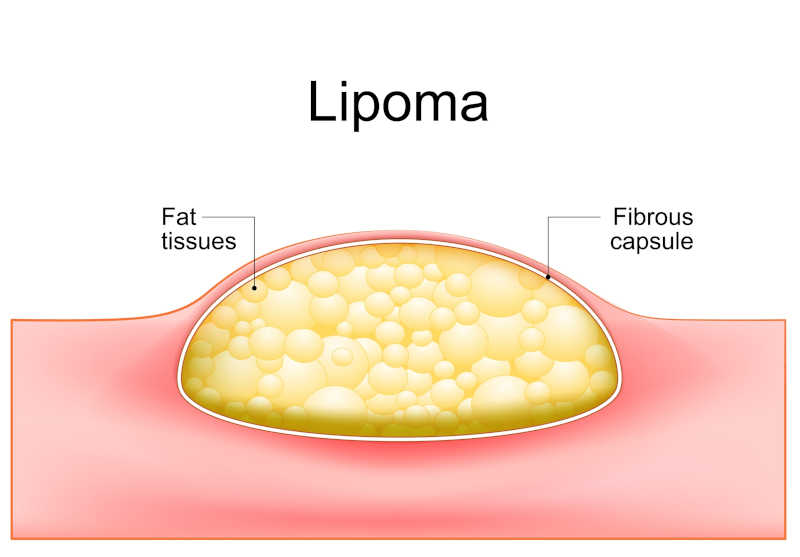
Lipomas are fatty tumors. True lipomas are non-cancerous and painless. They are typically soft and moveable. View more Lipoma pictures.
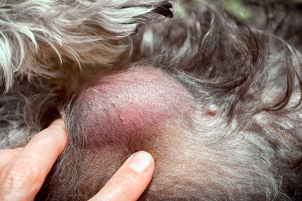
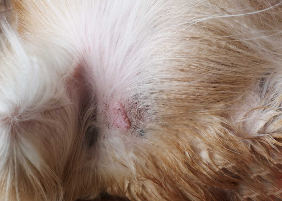
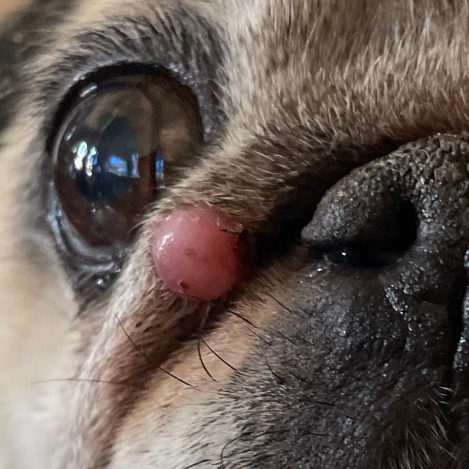
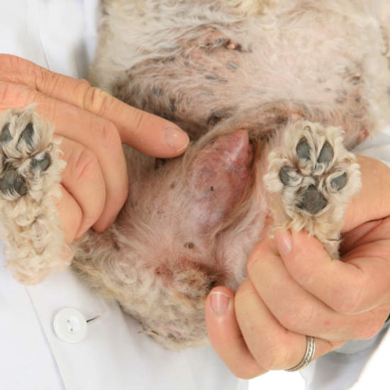
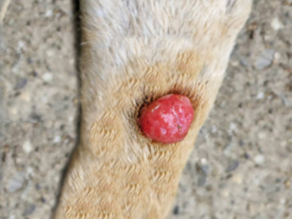
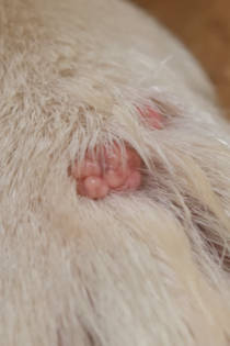
2. Sebaceous Cysts
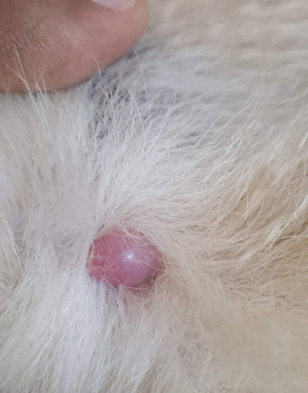
These masses appear as a fluid-filled sac and are non-cancerous. They are usually pink and hairless as shown in the two images below. View more pictures of sebaceous cysts.
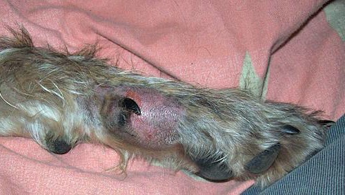
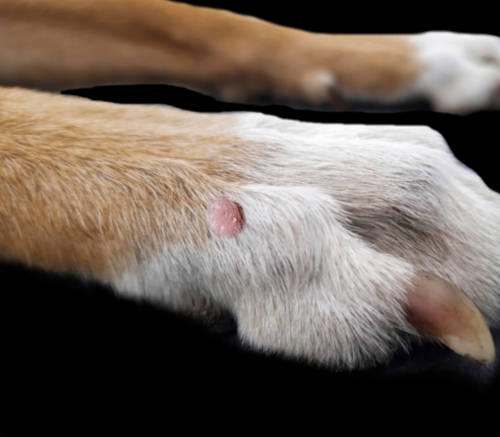
3. Interdigital Cysts
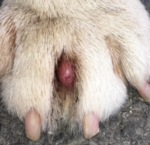
Interdigital cysts are found on the feet, between the toes. These lesions are areas of deep infection that form in response to chronic inflammation. They occur most commonly in larger, overweight dogs but can occur in any dog, especially if they are a result of a foreign body such as a sticker or grass awn in the paw. Learn more about interdigital cysts.
4. Mast Cell Tumors
These tumors can often be aggressive and appear white, pink, or covered in fur. View more mast cell tumor pictures.
5. Mammary Gland Tumors
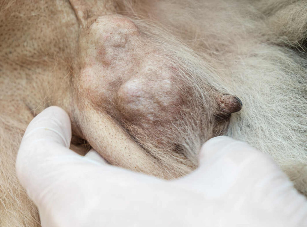
Mammary tumors can be cancerous or benign and are often next to or within the nipple. The mass may extend between multiple mammary glands and are typically firm. Mammary masses may have ulcerated skin overlaying them or be abscessed/bleeding. View more pictures of mammary gland tumors with veterinarian information.
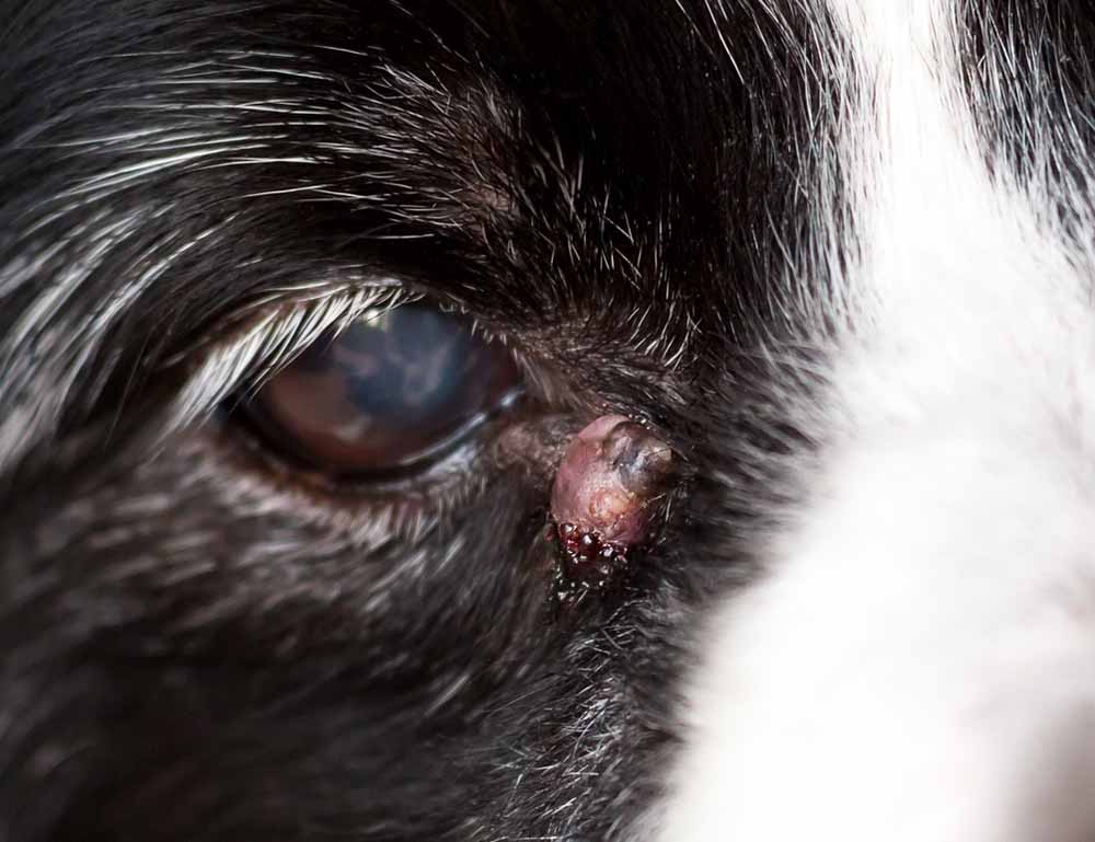
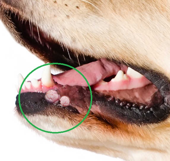
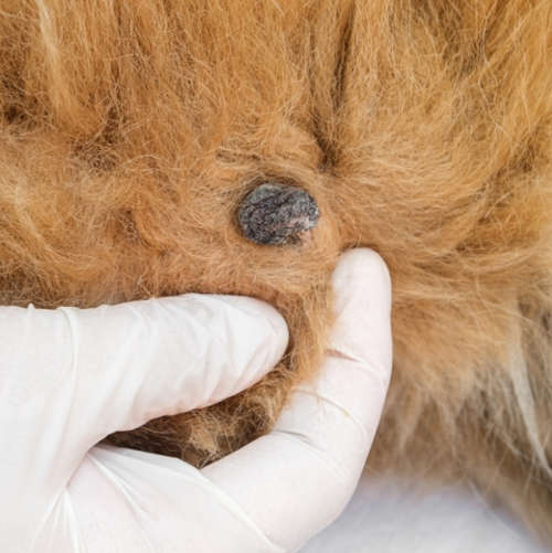
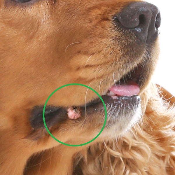
6. Melanomas
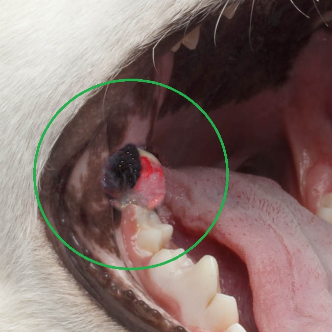
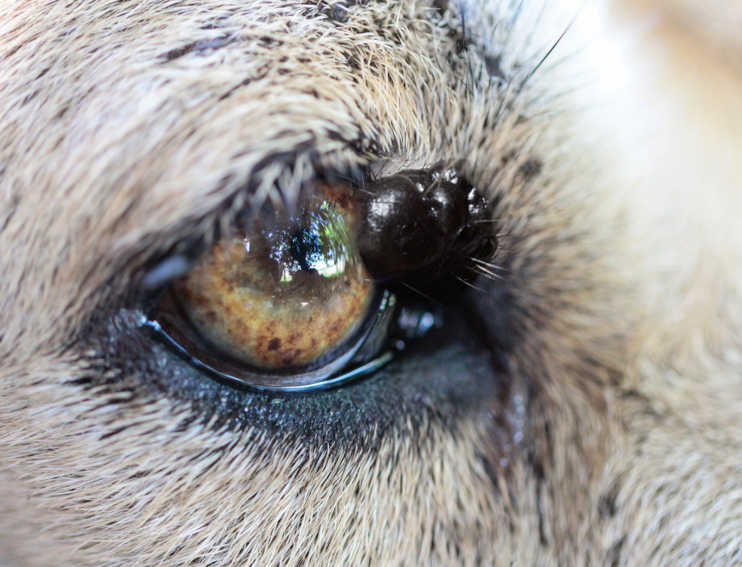
Melanomas may be cancerous or benign but are often very aggressive. These tumors are typically black, but not always and can be raised or flat. Above is a picture of a melanoma tumor in a dog’s mouth (black growth). Learn more about melanomas.
Please note that the pictures are provided as examples only. It usually is not possible to determine the real nature of a dog tumor, cyst, or growth by just looking at it. Learn about biopsies.
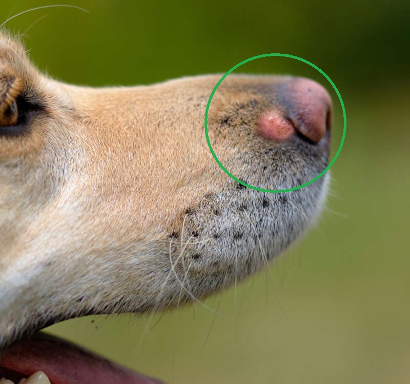
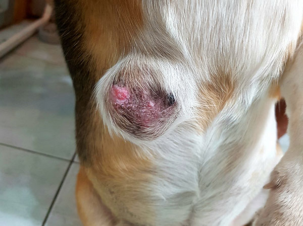
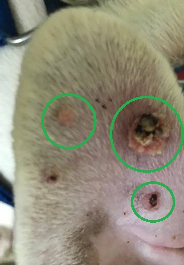
7. Squamous Cell Carcinomas
SCC is a malignant tumor that is typically raised and red. These are often found in light-skinned areas or in the mouth and may become ulcerated or bleed.
8. Soft Tissue Sarcoma
These masses are cancerous. There are many different types, depending on the tissue affected (including fibrosarcoma, hemangiosarcoma, liposarcoma). The masses can be soft or firm and are often invasive into surrounding tissue.
9. Anal Gland Tumor (Adenocarcinoma)
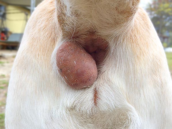
There are benign anal sac tumors, but adenocarcinoma are malignant. These appear as a growth on or just next to the dog’s anus.
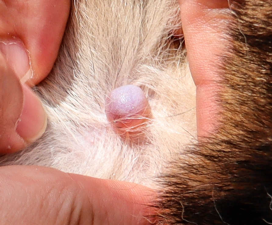
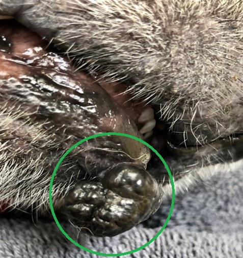
10. Warts
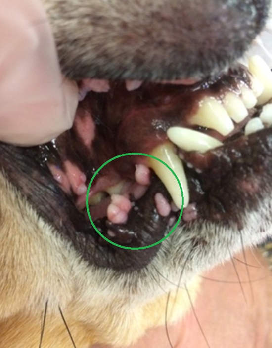
These lesions have a cauliflower-like appearance and can be single or a cluster of tiny lumps. The papilloma virus causes them and are benign. Warts often disappear after a few months. View more examples and pictures of warts or find out how to remove dog warts.
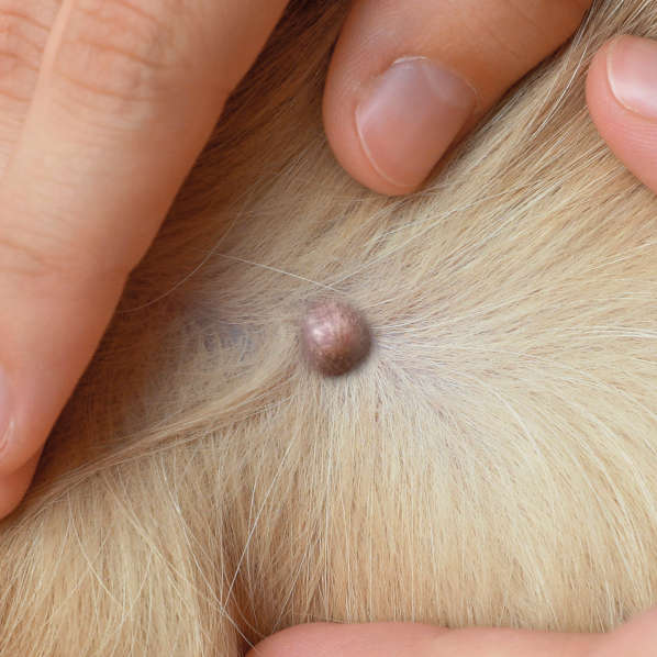
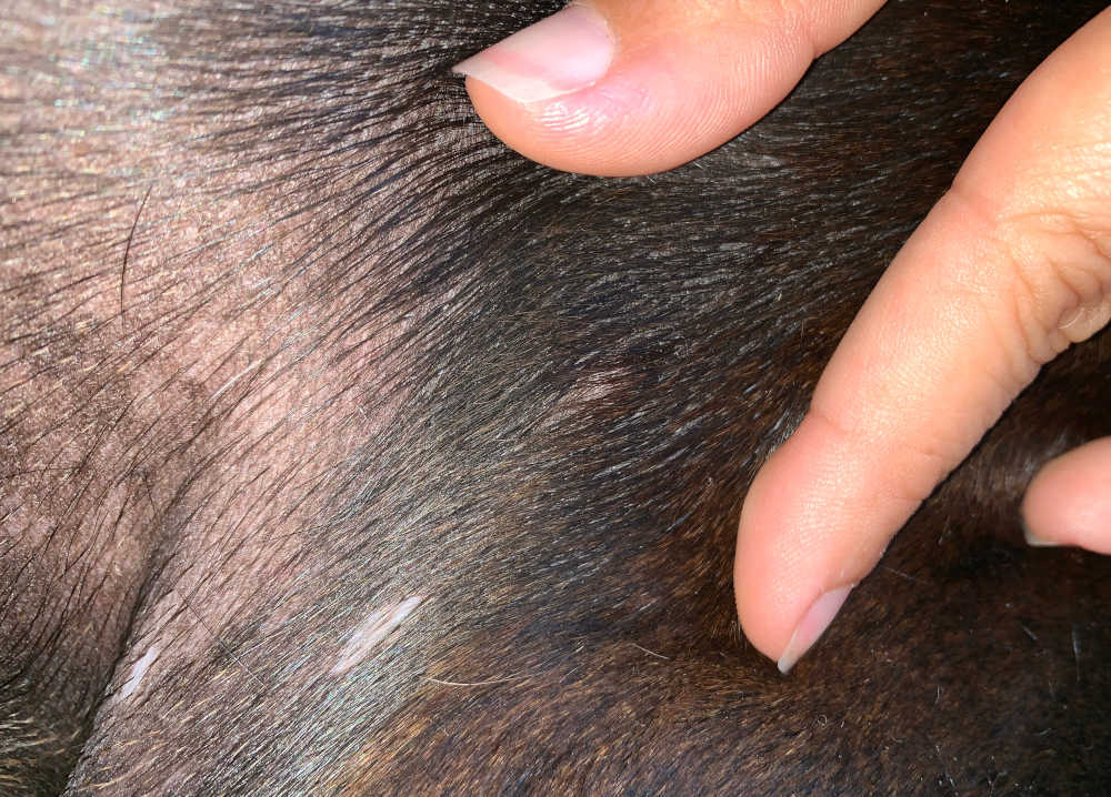
11. Skin Tags
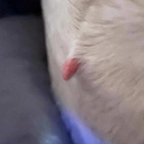
Skin tags are small, benign growths on the skin; are typically harmless and non-cancerous.
Learn more about Skin Tags.
Important: As veterinarians, we can never say what a dog’s growth is without testing it. For example, we have seen what looks like a lipoma actually come back as a malignant mast cell tumor. Always consult with our local veterinarian to confirm the diagnosis and determine the best treatment plan for your old dog.
Learn more:
- Go to our page on Lumps, Bumps and Growths for more information.
- View our page listing 21 skin conditions in dogs (with pictures).
- Learn more skin lesions or lumps due to cancer.
- Tumors commonly found on dogs’ legs.
Overview graphic:
Author
Disclaimer: This website's content is not a substitute for veterinary care. Always consult with your veterinarian for healthcare decisions. Read More.
My dog has developed about a dozen tiny lumps virtually over night. She also seemed to be under the weather. Does anyone have an idea?
.
Hives or some kind of rash/irritated reaction.
My dog has a few flesh colored lumps on his body. A new one appeared by his ear.
Any ideas?
I have a 9 year old English pointer. A year ago she started getting knots under her belly
. Over the year she has seen many vets. Now one know has gotten so big its draining all the time. They tried to do surgery because of her heart we about lost her the min they put her to sleep. They have tried 17 different medicines. They say it’s cancer and there’s nothing else that can be done. We are spending about $600.00 every 2 weeks on her. I just don’t know what to do!!!
My labrador had anal sack tumour removed over 3 years ago but just found out she has some more in her tummy she was on palladia for nearly 2 years then stopped it cost me over 23 thousand pounds at a Rough guess that’s the lower end
She is hoping going for a ct scan tomorrow see if she can be saved again my heart ❤️ is broken again
Sandra, I’m truly sorry your fur baby is going through this problem. First, please understand that I’m not a vet and not associated with animal medical conditions. As an animal lover with a fur baby, I am giving you a suggestion that I would do: First and foremost, is your pet in pain and/or does it interfere with his/her quality of life and Secondly, in the interest of your pet’s well being and doing what’s best…get a second opinion. I’m sorry I’m not much help. Wishing you all the best. Robin
I have a Mastiff and has a huge Knot on left side on top of his head, and a inch or so baove the left eye and goes all the way up and over the top towards the back of his head, is this a tumor or cancer?????♂️
I’m sorry to hear about your Mastiff’s new lump and appreciate your concern for him.
Cancerous lumps are often firmly attached to the skin beneath and tend to occur in older dogs.
Benign lumps such as cysts or fatty lumps can present in this way too.
We cannot say for sure what is going on without having him checked and running some tests such as a Fine needle aspirate or biopsy, to determine exactly what is going on.
If the lesion has been there more than a week and is growing or changing, we’d want to have it assessed in person.
“The information on this website is not a substitute for in-person veterinary care. Always seek advice from your veterinarian if you have concerns about your pet’s medical condition.”