

This article was updated on August 25th, 2023
If you are searching for information about blood blisters in dogs, you’ve likely noticed that a red, fluid-filled area has appeared on your dog’s skin somewhere. As veterinarians, we frequently see these red lumps or bumps, and their causes can range from very minor to malignant. In this article, I will explain the most likely causes (with pictures) and treatment options.
What’s a blood blister? is it serious?
A blood blister is technically an area of skin with a blood-filled fluid bubble, and it occurs in the outermost layer of the skin (the epidermis). Sometimes a blood blister can occur due to trauma. When this happens, it is likely benign and presents no real risk as long as the dog is not showing other signs of illness or pain.
In other cases, however, a blood blister can be due to a serious medical concern like cancer. Blood blisters should not be ignored.
“Sometimes a blood blister can occur due to trauma to the skin. These blisters are likely to go away on their own. However, any blood blister that persits for more than a few days should be examined by a veterinarian.”

Top causes of blood blisters (with pictures)
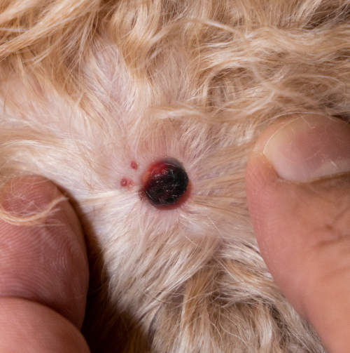
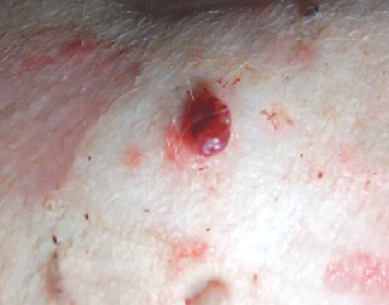
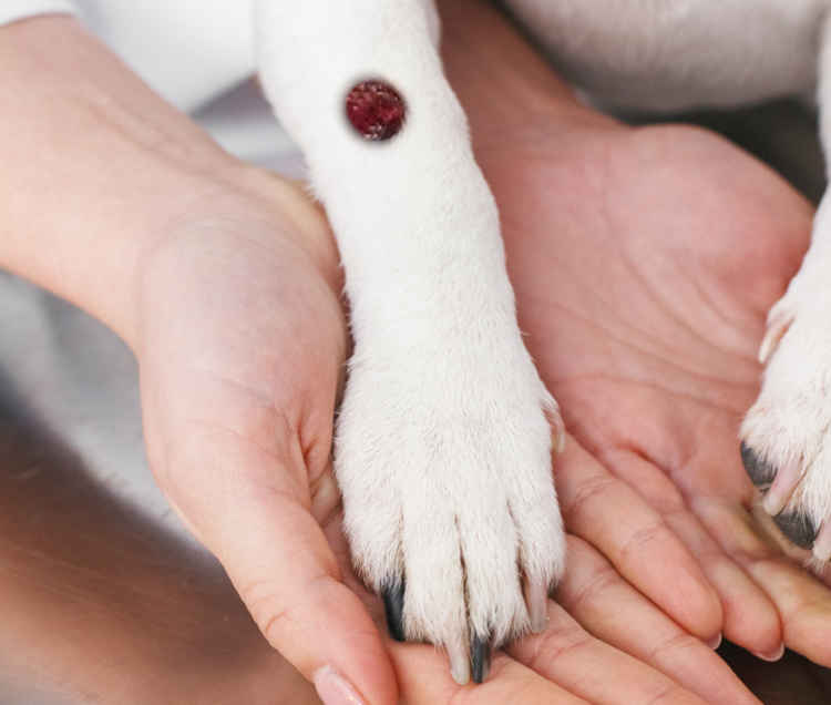
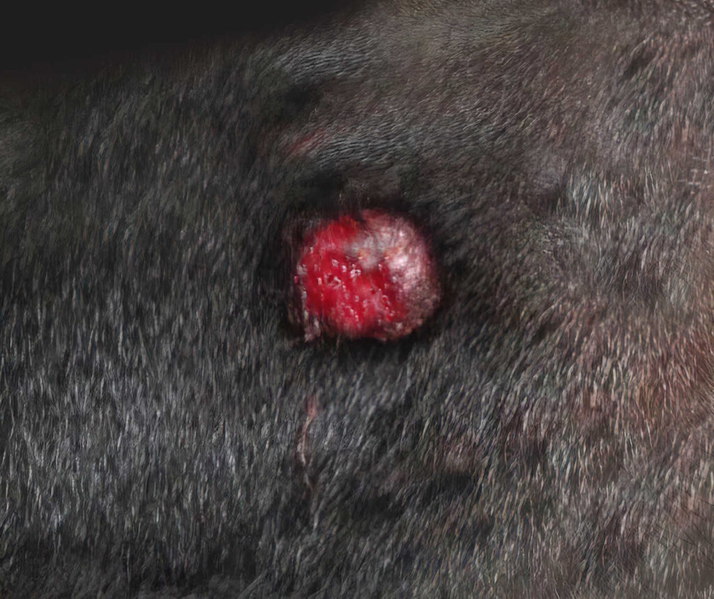
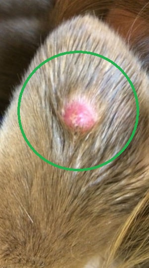
There are many types of skin lesions that look like “blood blisters”. They may look like a circular to oval lump or bubble, with red fluid within or a red color and bleeding around it. But they are not necessarily harmless blood blisters. Below are 5 conditions that can lead to blood blisters or red lumps:
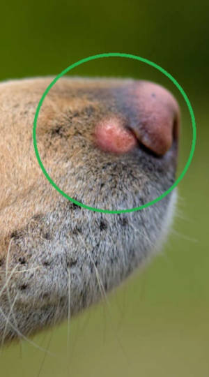
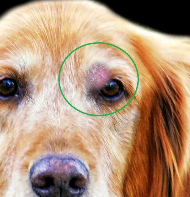
1. Mast cell tumors
According to research, mast cell tumors account for almost 18% of tumors found on dogs. They can take on a variety of appearances, but are often raised bumps with redness (or bleeding), as shown on the pictures below. Mast cell tumors can be diagnosed with fine needle aspirates, and due to the variable nature of tumor behavior, the chances for recovery vary significantly. Learn more about mast cell tumors with pictures.
2. Cutaneous hemangiosarcoma
These masses are typically small and occur on a dog’s belly, on the outside layer of skin. They are usually not as quick to spread as other types of hemangiosarcoma. However, because they are cancerous, they still require surgery for removal, and sometimes additional treatments like chemotherapy.
3. Hemangioma
Hemangiomas are a benign cancer and can cause dark purple blisters on the skin. They arise from blood vessels and UV radiation is thought to play a role in in the disease. Dogs with short fur and light skin are more likely to get these, especially in areas of the body with less hair that’s exposed to sunlight, such as the chest and belly. Surgical removal is usually recommended.
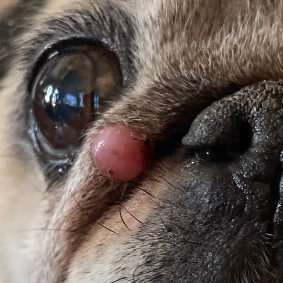
4. histiocytomas
Histiocytomas make up 15.9% of tumors found in dogs. They are small benign lumps that appear most commonly in young dogs (under three years old). Histiocytomas form when immune cells called Langerhans cells over-react and replicate rapidly. Treatment is simple. In most dogs, histiocytomas regress (shrink) in 3 months. In rare cases, a dog may have multiple histiocytomas. Multiple histiocytomas are a symptom of a dangerous skin disease called histiocytosis. In histiocytosis, affected dogs have multiple lumps on their body.
IMPORTANT: It is usually not possible to diagnose your dog’s lump or blister based on its appearance alone. Veterinarian diagnosis may be required to determine the right treatment.
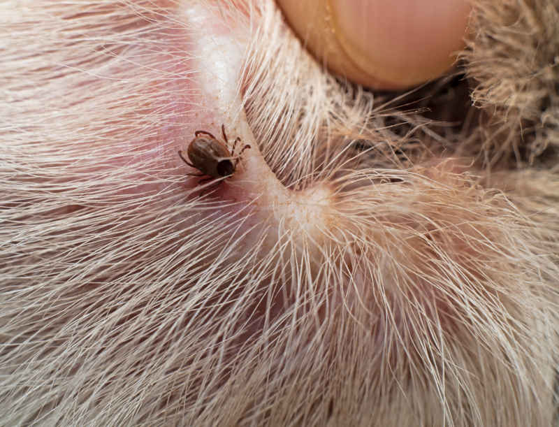
5. Ticks
Sometimes an engorged, feeding tick can look like a blood blister.
When should you call your veterinarian
If you notice an area or a lump on your dog’s skin that appears red, has blood within or is bleeding, you should make an appointment with your vet, especially if it has not resolved after a few days.
As discussed above, these lesions can be caused by a variety of different conditions and are typically hard for even a vet to diagnose just by looking at them.
A word about ear hematomas
If your dog has a swollen ear or a firm, fluid-filled sac in the ear region, it is possible they may have an ear hematoma.
If so, it is not a life-threatening problem but is usually uncomfortable, so call your vet and try to have your dog seen within a few days if possible. If there is active bleeding that you can see, this becomes an emergency, so bring your dog to the vet immediately.
If your dog has any of the above abnormalities but is not eating, is lethargic, seems painful, or is not acting like their normal selves, it is always advised to have a vet evaluate them urgently.
What you can do at home while waiting for a vet appointment
For other skin problems that look like blood blisters:
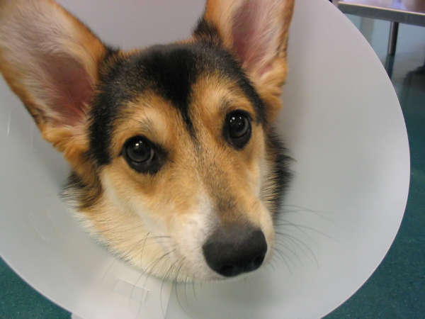
1. Prevent your dog from damaging the area. An e-collar or loose t-shirt covering it can prevent your dog from licking and scratching and causing more damage to the lesions.
2. Monitor the affected area once or twice daily and note any changes in size, shape, or color. Taking photos to show the vet can be helpful, especially if the area is changing in appearance.
3. Clean the area and keep it dry. For small amounts of oozing blood, very gently keep the area clean and dry.
If there is a large amount of active bleeding, take your dog to the emergency vet.
Learn more about common dog skin issues and diseases.
Ear hematomas in dogs: What do they look like, and why do they occur?
A hematoma is the leakage of blood from blood vessels into the surrounding area. This can occur on the surface of the body or internally, where it is not visible. This commonly occurs due to injury such as excessive force. Less commonly, blood clotting disorders may prevent the body’s normal process of stopping blood flow after an injury and lead to bleeding or large hematomas.
Common locations of hematomas:
- Ears – occurs when a dog shakes their head violently and breaks fragile blood vessels in the ear flap. Read our article about ear hematomas.
- Neck or limbs – occasionally, a dog can get a hematoma at the vet if they have trouble staying still during a blood draw. These are usually small, swollen areas near the underside of the neck or on the limbs and will resolve on their own.
- Scrotum – hematomas in the scrotum (the sac around the testicles) can form after a neuter surgery due to excessive movement during tissue healing.
- Internally – these are typically not visible to a dog owner but can occur in organs with high blood flow, such as the spleen.
Appearance
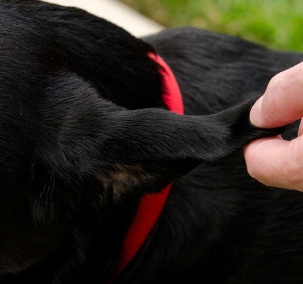
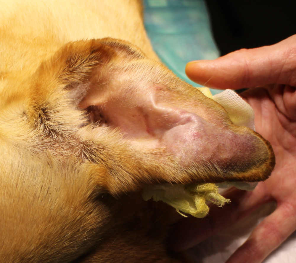
Hematomas can vary greatly in size and appear as swollen lumps underneath the skin. Hematomas of the ear can cause the whole ear to swell and feel very firm, warm, and painful. From the side, the dog’s ear will look thickened (see below) to almost bursting like a balloon compared to the normally thin ear flap.
Photo of dog ear hematoma from the side:
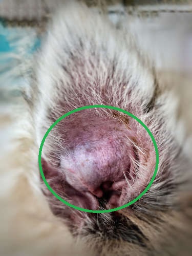
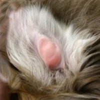
Smaller ear hematomas, or the beginning of one, can look like a small bulging, raised area on the inside of the ear flap, such as in this photo below:
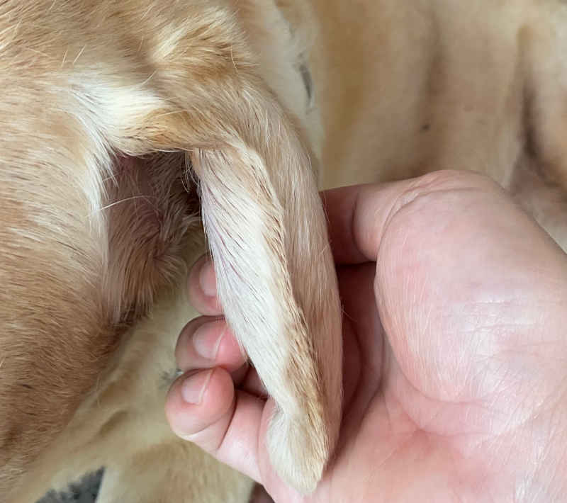
Another photo of an ear hematoma:
Ear hematomas are a very common problem in dogs. They occur due to trauma or excessive movement, such as a dog violently shaking their head from an itchy ear infection or something in their ear, like a grass awn. Sometimes dog bites that don’t break the skin can cause ear hematomas. Ear hematomas can occur in any dog breed but are more common in dogs with floppy ears and certain breeds predisposed to allergies and ear infections, such as Labradors, Golden Retrievers, and Pit Bulls.
The veterinary visit: what to expect for diagnosis, treatment, outcome
Diagnosis
Diagnosis of a suspected ear hematoma is straightforward. The vet will palpate and examine the swollen ear and the ear canal. Testing for an ear infection is usually recommended to treat the underlying reason causing the dog to shake its head.
For skin abnormalities that look like blood blisters, diagnosis may involve a monitor and follow-up approach if the lesion appears benign, or testing such as a fine needle aspirate or skin biopsy. These tests can help to identify potential tumors or other conditions. If your dog has other symptoms or is not feeling well, bloodwork, chest x-rays, and an abdominal ultrasound may be recommended.
Treatment
For ear hematomas: The goals of treatment are to prevent further bleeding in the ear flap and resolve the underlying problem that’s causing the dog to shake its head in the first place. Treatment of ear infections or skin allergies is often required. If the hematoma is large or causing discomfort, the vet may offer drainage of the hematoma and injection of a medication into it, or surgery to eliminate the space between the skin and the cartilage to prevent the hematoma from recurring. For home care, there are specific bandages made to prevent movement of the ear, or oral sedatives to decrease your dog’s activity during recovery.
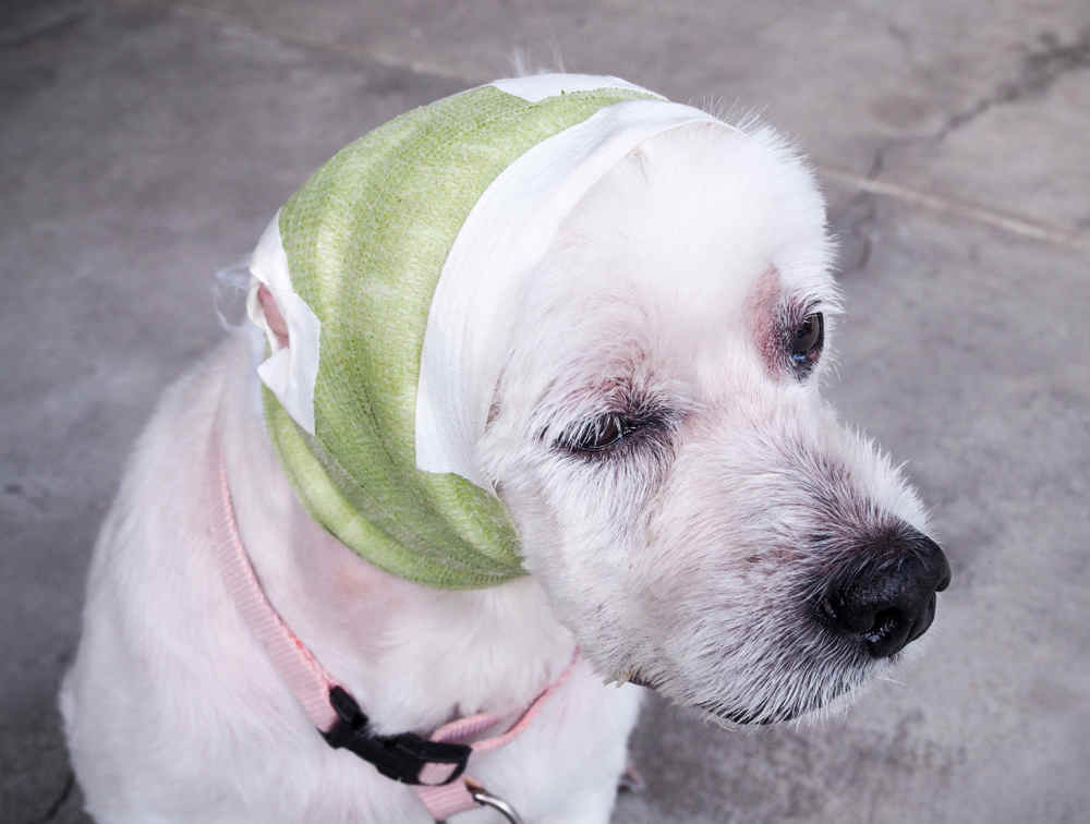
Example of a bandage for an ear hematoma:
Management of recurring ear hematomas can be frustrating due to cost and difficulty keeping dogs comfortable. Treatment with one option may not always be successful, and the vet may have to recommend an alternative. Occasionally, if no treatment is working well, or due to financial constraints, owners may decide against treatment. This is not recommended, as it can take several weeks of a dog being in pain before it will heal and will lead to scarring and deformation of the ear.
For other skin lesions, treatment will heavily depend on diagnostic test results. If there is concern for a serious problem such as cancer, treatment can involve surgery and sometimes chemotherapy.
How to prevent ear hematomas in the future
Prevention of ear hematomas usually involves the prevention of the underlying problem that causes a dog to shake their head and ears excessively. The most common cause is an ear infection or underlying skin allergies.
Excessive shaking of the head like this can cause ear hematomas:

Dogs that are beginning to develop an ear infection often scratch more than normal, shake their heads frequently, and have a foul odor from the ears. If you notice these behaviors, contact your dog’s regular vet for an appointment as soon as possible, before the problem gets more severe.
Although it is not usually a life-threatening condition, an untreated ear infection can lead to ear hematomas, which are significantly more costly and time-consuming to treat. It is not recommended to try home treatments for cleaning ears without consulting a vet, as it can make the problem worse.
Related:
Pictures of 21 common skin problems in dogs.
Author
Disclaimer: This website's content is not a substitute for veterinary care. Always consult with your veterinarian for healthcare decisions. Read More.