
You might have noticed that your dog’s ear feels puffy and filled with fluid, unlike the soft and smooth ear it used to be. Your dog might also be scratching their ears or shaking their heads, and you are wondering if your dog has an ear hematoma. To help, our veterinarian Dr. Linda Simon has put together this quick visual guide to show you what ear hematomas look like.
What an Ear Hematoma Looks Like

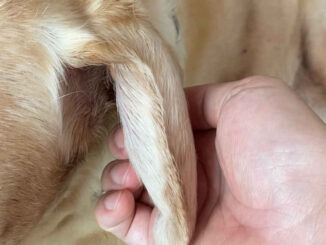
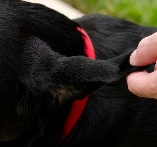
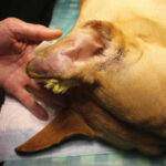
If your dog has a hematoma, the ear will appear swollen and doughy, almost like a thick water balloon. It has this appearance because it is a blood-filled swelling between the skin and cartilage layers of the earflap. It can affect the whole earflap or just part of it. In the picture below we can see a hematoma at the base of the ear pinna:
In the picture above, we can see that the skin is a bit ‘leathery looking’ and red with small scratches on the surface. In this instance, the dog likely has an underlying ear infection. This will have caused shaking and scratching which will have led to blood vessels bursting within the ear flap, and the hematoma forming.
Ear hematomas: a visual gallery
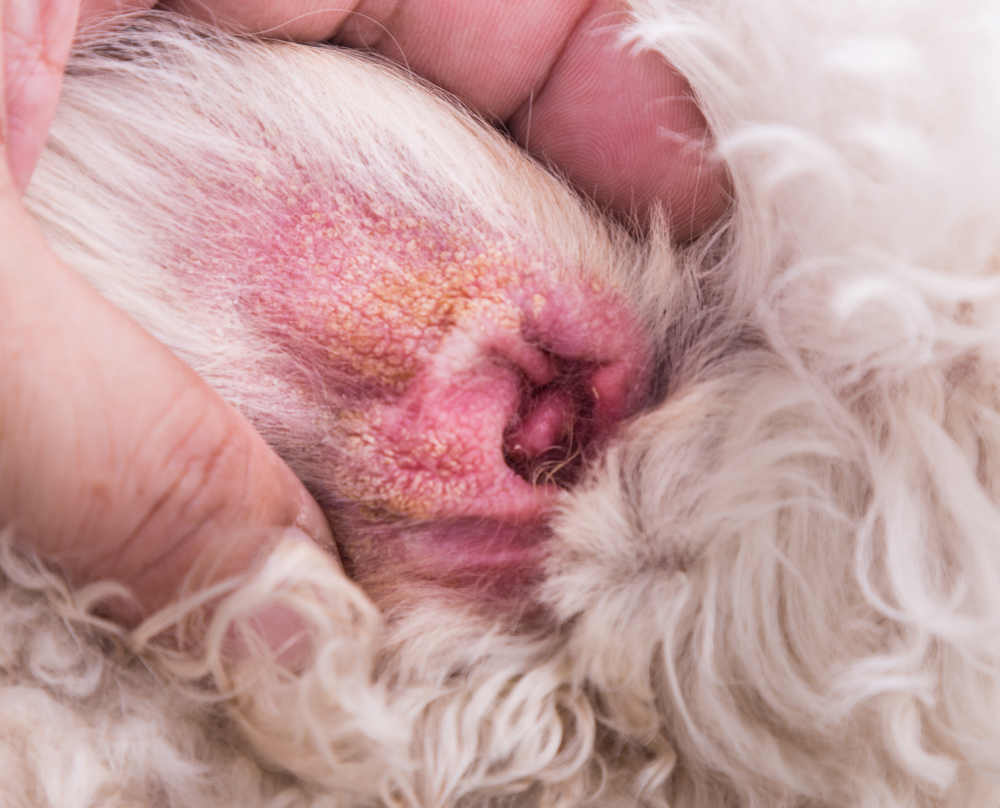
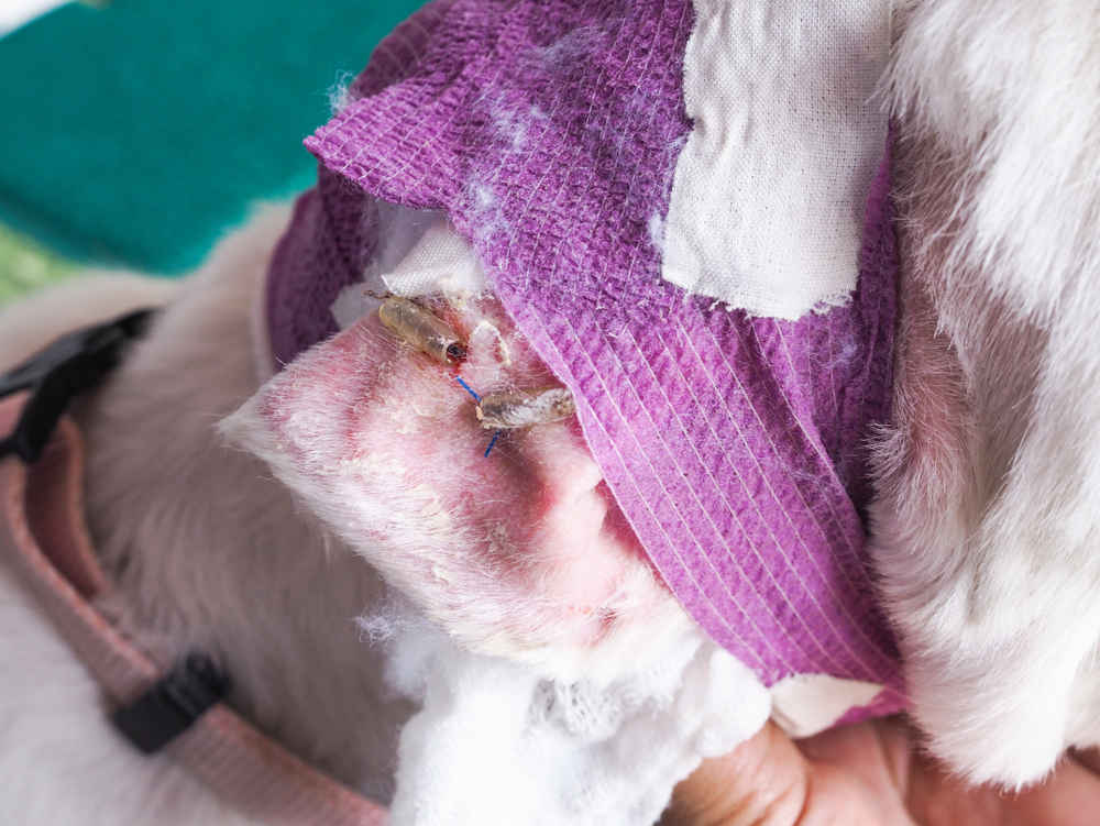
The white ear below is acutely infected. The skin is bright red and swollen and we can also see plaques, or thick scales/crusts. For many dogs, infections like this are linked to underlying medical issues such as allergies or atopic dermatitis. When an ear is infected, it is more likely a hematoma will form.
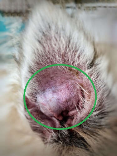
The black ear above is puffy and swollen, indicating an aural hematoma. We would be wondering why this has occured, and should be checking for any allergies, infections or ear mites.
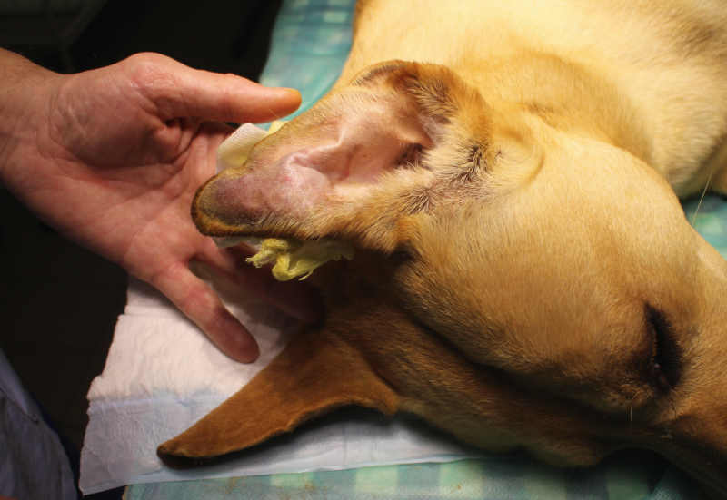
In the picture above with the beige-furred dog, we can see that a hematoma has formed at the top of the ear pinna. The puffiness is caused by excess blood within the pinna. This will be a combination of clotted and non-clotted blood. The ear will generally feel warm and will be tender for the dog when touched.
Key characteristics
You may initially notice your dog scratching their ears or shaking their head excessively. Some dogs will even tilt their head to one side when one of their ears bothers them. If you see any of these symptoms, keep a close watch on the earflaps for any of the following:
- Swelling in the ear flap: The ear flap appears puffy and swollen due to the accumulation of blood inside.
- Warmth to the touch: The swollen area feels warmer compared to other parts of the ear or body.
- Redness: The skin over the hematoma may look redder than usual, indicating inflammation.
- Pain or discomfort: The dog may show signs of pain, like whining or shaking its head, when the area is touched.
- Altered shape of the ear: The ear may look misshapen or droop differently due to the swelling from the hematoma.
- Smell: Oftentimes the affected ear is infected, so the owner may notice a foul or musty smell.
Medical interventions
There are a range of treatment options, and the route taken will depend on a number of factors including the size of the hematoma, how long it has been present and the age and general health status of the patient. Finances also have to be considered, as the dog may need to be put under general anesthetic for more invasive procedures.
It is crucial that any underlying ear infections or allergic flare ups are treated, rather than only focusing on treating the hematoma itself. This may mean a swab is taken, to determine what type of infection is present and which medicine would be indicated.
Simple aural hematomas may be treated by inserting a needle to drain the blood. The vet may also locally inject some steroid, in an effort to prevent the pinna from re-filling.
When this is not successful, the ear flaps may need to be temporarily tacked together for a couple of weeks. This can be done with e.g. sutures, buttons or the plastic pieces of a fluid drip. We want the flap to heal together, without filling with fluid such as blood.
Learn more about Treatment Options.
Home care and recovery
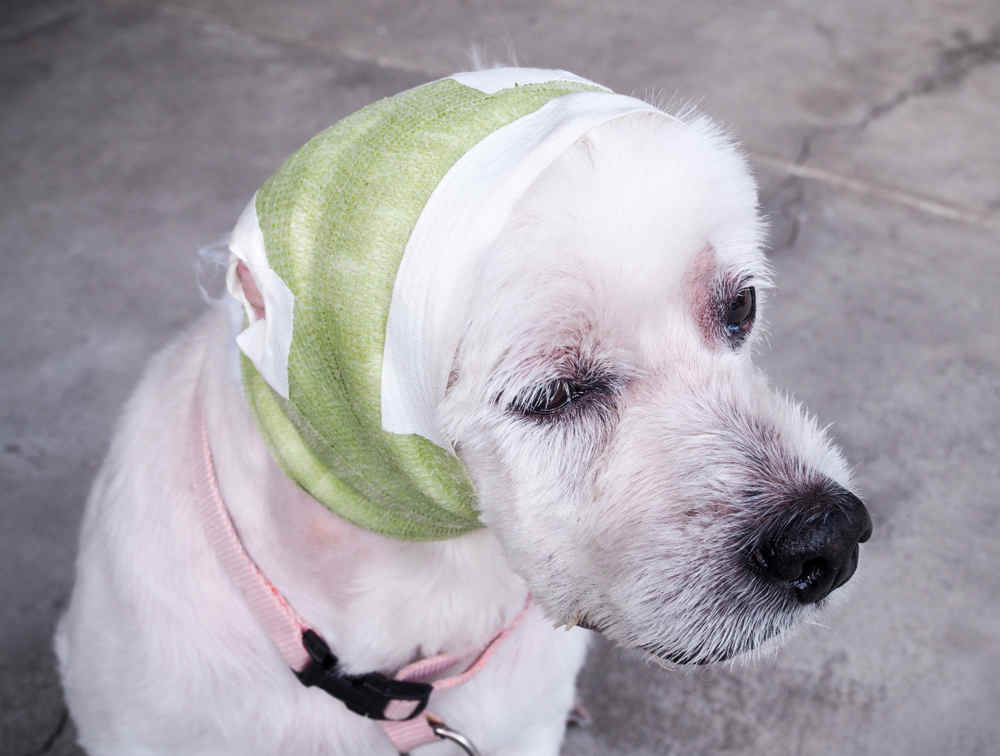
After the procedure, we want the dog to keep their head still as any shaking or rubbing at the ears can lead to the hematoma forming again. Oftentimes, the ear will be bandaged up on top of the head, so it is immobile. The dog may also have a buster collar, to prevent the bandage being pulled off.
Your vet may issue some medicine such as pain relief, anti inflammatories and antibiotics, and this medication should be given as prescribed.
Your vet will likely ask to see your dog back for a couple of check ups over the coming weeks. Usually after two weeks, any sutures or other material would be removed.
Learn more about Dog Ear Hematoma Wraps.
Related posts:
 Cauliflower Ear in Dogs: What is It & How to Help Your Dog - Cauliflower Ear in Dogs is a common condition that can affect many breeds and results in great discomfort for your… [...]
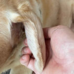 Home Remedies to Help Dogs with Ear Hematomas, by Dr Guise - You’ve found a crazy-looking lump on your dog’s ear and are wondering what you can do to help your poor… [...] Dog Ear Hematomas: A Dog Owner’s Guide - It can be alarming to rub your dog’s ear and feel a puffy, fluid-filled ear that once was soft and… [...]
 My Dog’s Ear Hematoma Popped: What Should I Do? - In my practice, we see dogs with ear hematomas at least once a month or more. It is incredibly rare… [...]
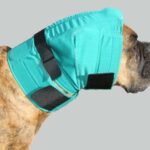 Dog Ear Hematoma Wraps: A Dog Owner’s Guide - Image credit: No Flap Ear Wrap Manufacturer. Overnight your dog’s ear ballooned up into a hot, squishy mess known as… [...]
Author
Disclaimer: This website's content is not a substitute for veterinary care. Always consult with your veterinarian for healthcare decisions. Read More.