
This article was updated on October 1st, 2023
As a veterinary ophthalmologist with extensive experience, I have successfully treated many cases of third eyelid conditions in dogs.
In this article, I’ll provide a comprehensive overview of what the third eyelid in dogs is and the associated conditions. This information will help you promptly identify and manage these conditions to ensure your dog’s well-being and avoid any complications with their eyes.
What is a third eyelid in dogs?
The third eyelid (or nictitating membrane) is a mobile, protective, immunologic, and glandular structure lying between the eyeball and the lower eyelid((Slatter’s Fundamentals of Veterinary Ophthalmology; 5th edition; David J. Maggs; Paul E. Miller; Ron Ofri; Third Eyelid; Chapter 8; page 159 – 164)).
Important functions of the third eyelid
The third eyelid is essential in maintaining eye health. It performs functions like:
• Distributing of the precorneal tear film, which is the thin layer of tears that covers the surface of the eye just before it reaches the cornea
• Protecting the cornea
• Producing part of the aqueous component (the watery layer) and immunoglobulins (antibodies) of the tear film((Slatter’s Fundamentals of Veterinary Ophthalmology; 5th edition; David J. Maggs; Paul E. Miller; Ron Ofri; Third Eyelid; Chapter 8; page 159 – 164))

Diagram of the eye showing normal position of the third eyelid((Modified from Evans HE: Miller’s Anatomy of the Dog, ed 3, Philadelphia, 1993, Saunders: Slatter’s Fundamentals of Veterinary Ophthalmology; 5th edition; David J. Maggs; Paul E. Miller; Ron Ofri; Third Eyelid; Chapter 8; page 159 – 164))
Clinical symptoms of an issue with the third eyelid
While it’s not always obvious when there’s a problem with the third eyelid, there are certain signs and symptoms that can indicate an issue:
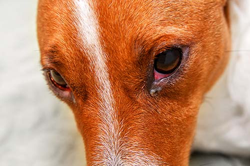
- Redness of the conjunctiva, the transparent mucous membrane that lines the inner surface of the eyelids
- Conjunctival swelling
- Discharge from the eye
- Third eyelid protrusion
- Frequent squinting
Main issues causing third eyelids in dogs
In this section, we’ll outline the primary causes of third eyelid issues in dogs. Understanding these potential factors is essential for recognizing and addressing eye problems in our canine companions.
Cherry eye (third eyelid gland prolapse)
Prolapse of the gland of the third eyelid (commonly referred to as cherry eye) occurs most commonly in dogs and occasionally in cats((Slatter’s Fundamentals of Veterinary Ophthalmology; 5th edition; David J. Maggs; Paul E. Miller; Ron Ofri; Third Eyelid; Chapter 8; page 159 – 164)).
The clinical signs
The following symptoms may indicate Cherry Eyes:
- Red-pink mass behind the third eyelid
- Ocular discharge
- Squinting
- Corneal ulceration
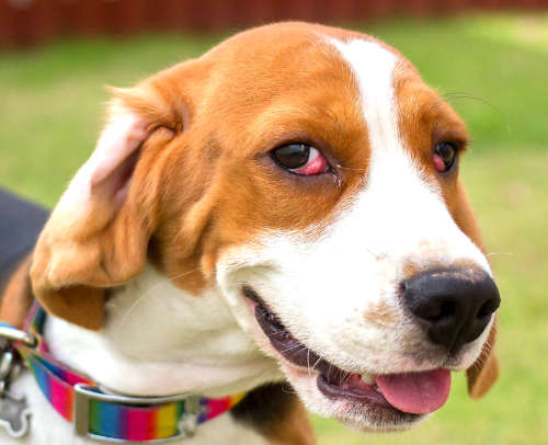
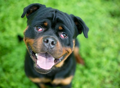
Prolapse of the gland of the third eyelid (cherry eye) in a dog((Slatter’s Fundamentals of Veterinary Ophthalmology; 5th edition; David J. Maggs; Paul E. Miller; Ron Ofri; Third Eyelid; Chapter 8; page 159 – 164))
The cause of Cherry Eyes
This condition is often observed in young animals who are exposed to environmental antigens for the first time. It is believed to be caused by a combination of inflammation in the gland and a weakening of the ligament that connects the third eyelid gland to the bone.
Breeds that have a shortened skull, like Pugs, are more susceptible to a weakened ligament. This unique combination of factors enables the gland to protrude while still being attached to the cartilage of the third eyelid((Slatter’s Fundamentals of Veterinary Ophthalmology; 5th edition; David J. Maggs; Paul E. Miller; Ron Ofri; Third Eyelid; Chapter 8; page 159 – 164)).
Treatment of a Cherry Eye
It’s important to replace the gland through surgery as soon as possible to maintain function and prevent dryness, inflammation, secondary infection, and an unpleasing appearance of the exposed gland and conjunctiva. Swift surgical intervention will help preserve the overall health and aesthetics of the affected area.
It’s notable that keratoconjunctivitis sicca, an inflammatory condition of the cornea and conjunctiva, is commonly seen days to years later in animals, especially those of susceptible breeds in which the third eyelid or its gland was removed. (Slatter’s Fundamentals of Veterinary Ophthalmology; 5th edition; David J. Maggs; Paul E. Miller; Ron Ofri; Third Eyelid; Chapter 8; page 159 – 164)
When to contact your vet
Contact your vet when you notice a problem with your dog’s eyes. Eyes are precious, and the problems could get worse if they aren’t treated quickly.
Tumor affecting the third eyelid
Different types of primary tumors can affect the third eyelid.
If a third eyelid neoplasm (tumor) is identified, it’s important to conduct a comprehensive evaluation of the orbit, regional lymph nodes, and other distant sites to check for potential spread of the tumor or metastases. This thorough assessment will help determine the extent and staging of the cancer.
Treatment
Surgical excision is recommended for all malignant tumors other than lymphoma, which can be treated via systemic chemotherapy.
For focal masses near the free margin of the third eyelid, resection of the mass and a margin of surrounding normal tissue may be possible. Larger tumors necessitate complete excision of the third eyelid and surrounding conjunctiva((Slatter’s Fundamentals of Veterinary Ophthalmology; 5th edition; David J. Maggs; Paul E. Miller; Ron Ofri; Third Eyelid; Chapter 8; page 159 – 164)).
When to contact your vet
It is crucial to reach out to your veterinarian if you notice any issues with your dog’s eyes. Their eye health should not be taken lightly, as delays in treatment could potentially lead to worsening conditions. Prompt communication with a vet will ensure that appropriate measures are taken to address the problem effectively.
Trauma to the third eyelid
The third eyelid, also known as the nictitating membrane, can sustain injuries for several reasons. These include road traffic accidents, cat fights, or the presence of a foreign object in the eye. It’s important to be aware of these potential causes and take appropriate measures to prevent or address such injuries.
If the tear only involves the conjunctiva, it typically does not need stitching. In cases where there are small flaps on the leading edge, they can be safely removed.
Larger lacerations, especially those involving the free margin and creating larger loose flaps, usually should be carefully debrided and apposed, ensuring that the cartilage is well covered by conjunctiva.
Although some retraction takes place during healing, a functional third eyelid is typically retained. In some circumstances grafting of oral mucous membrane may be useful for replacing large defects((Slatter’s Fundamentals of Veterinary Ophthalmology; 5th edition; David J. Maggs; Paul E. Miller; Ron Ofri; Third Eyelid; Chapter 8; page 159 – 164)).
When to contact your vet
If you observe any problems with your dog’s eyes, it is essential to promptly contact your veterinarian. Taking the health of their eyes seriously is crucial because delaying treatment could result in worsening conditions. By reaching out to your vet right away, you can ensure that appropriate measures are taken to address any issues and prevent further complications.
Conjunctivitis
Conjunctivitis refers to the inflammation of the conjunctiva, a delicate tissue layer that covers the inner eyelid and extends across the surface of the eye. This condition can arise from various causes.
Symptoms of Conjunctivitis
Common signs for one or both eyes include:
- Red and swollen eye
- Squinting more or closing eyes
- Change of corneal color
- Scratching eyes or face
- Not eating
- Lethargy
Causes of conjunctivitis
These are some (but not all) of the underlying causes that contribute to conjunctivitis:
- Bacterial or viral infections
- Dry eye
- Eyelid or eyelash issues
- Allergies
- Irritants
- Corneal ulcers
- Glaucoma (increased pressure inside the eye)
- Foreign bodies
- Uveitis (inflammation inside the eye)
Treatment
During the preliminary assessment, your veterinarian will conduct a comprehensive examination of your dog’s eyes in order to identify the underlying cause. This step is crucial in determining the appropriate treatment required for your beloved pet.
When to contact your vet
Should you observe any signs of an eye problem in your canine companion, it is crucial to promptly reach out to your veterinarian. Taking immediate action can help prevent the condition from worsening swiftly.
Horner syndrome
Horner’s syndrome refers to a widely recognized neurological disorder that specifically affects the third eyelid, as well as the eye and facial muscles. This condition may present itself suddenly and predominantly impacts one side of the head. However, there are rare instances in which it can have bilateral effects on both sides.
Common signs of Horner Syndrome
Horner’s syndrome symptoms in dogs are not always obvious and may include:
- Protrusion of the third eyelid
- Upper eyelid ptosis (drooping of the upper eyelid)
- Pupil constricted
- Enophthalmia (globe appears sunken)
Common causes of Horner Syndrome
Horner’s syndrome is due to a dysfunction of the sympathetic nerves of the eyes and surrounding facial muscles. This is part of the autonomic nervous system, which helps to control normal functions such as blinking, muscle tone, etc.
Some of the most common causes include:
- Puncture wound
- Infection
- Middle or inner ear diseases
- Tumor
- Intervertebral disc disease
- Fracture skull
- Injury to the back of the globe
- Idiopathic refers to a condition or phenomenon that lacks a known or identifiable cause.
Treatment
Some of Horner’s syndrome will resolve spontaneously; however, it is important to treat any underlying disease. There are several diagnostic tests that will be performed to determine if there is an underlying cause in your pet including an eye and ear exam, radiographs (X-rays) of the skull and chest, and possibly advanced imaging such as CT scans or MRIs. Pharmacologic tests may include phenylephrine drops placed in the affected eye to help localize the source of the problem.
When to contact your vet
If you happen to observe any indications of eye issues in your beloved pet, it is of utmost importance to promptly reach out to your veterinarian. Taking action can play a vital role in preventing the condition from worsening.
Author
Disclaimer: This website's content is not a substitute for veterinary care. Always consult with your veterinarian for healthcare decisions. Read More.